NUTRITION, DENTITION AND DIGESTION IN MAMMALS
Objectives
- Explain the concept Nutrition.
- Classify nutrients found in food.
- Demonstrate the presence of various nutrients found in food.
- Explain the importance of dental care in humans.
- Explain the concept of balanced diet and malnutrition.
- Explain the concept enzymes.
- Describe the process of digestion and absorption
- Describe the structure and functions of the liver.
NUTRITION IN MAMMALS
Nutrition is the process by which organisms make or obtain food and utilizing it for growth maintenance. There are two types of nutrition. These are heterotrophic and autotrophic (holophytic) nutrition.
Food
Food is any substance consumed to provide nutritional support for the body. Food contains essential nutrients, such as carbohydrates, fats, proteins, vitamins, water or minerals. The substance is ingested by an organism and assimilated by the body cells to produce energy, maintain life, or stimulate growth.
Why Living Organisms Require Energy
2. Electrical transmission of nerve impulses
3. Synthesis of substances, such as proteins
4. For transport of molecules against concentration gradients
Carbohydrate
A carbohydrate is an organic compound that consists of only carbon, hydrogen, and oxygen. The hydrogen and oxygen atoms are present in the ratio 2:1 (as in water). The general formula for carbohydrates is Cm(H2O)n, where m and n are whole numbers.
Source of carbohydrates:
Class of Carbohydrates
Three main groups of carbohydrates are monosaccharide, disaccharides, and polysaccharides.
Monosaccharides
Monosaccharides mono, means "one", and saccharide, means "sugar". The general chemical formula is CnH2nOn; where n is three or more carbon atoms.
Monosaccharides with three carbon atoms (C3H6O3) are called trioses, those with four (C4H8O4) are called tetroses, five are called pentoses, six are hexoses, and so on.
Hexose sugar: there are three types of hexose sugar; glucose (dextrose), fructose,
and galactose.
They have the same general formula C6H12O6, but different arrangement of atoms. They are the simplest form of sugar which the body absorb and assimilate. They are the building blocks (monomers) of disaccharides and polysaccharides.
Monosaccharides are usually whitish, crystalline solids, water-soluble, sweet taste and reducing sugar. They are reducing sugars because they reduce blue copper (II) ions to brick red copper (I) ions. i.e. cupric oxide to cuprous oxide.
Disaccharides
Disaccharides contain two sugar units i.e. two monosaccharide units bound together by a condensation reaction or dehydration reaction.
Polysaccharides
Polysaccharides are polymers formed by condensation of several monosaccharide units.
Disaccharides and polysaccharides are broken down by hydrolysis into
monosaccharides.
Functions of Carbohydrates
□ Provide energy for cell activities
□ Form structural components in plants and arthropods (cellulose, lignin, pectin).
□ Form components of coenzyme and genetic molecule
□ Take part in biological transport, cell-cell recognition, activation of growth factors.
□ Required in biosynthesis of proteins and lipids.
Test for Carbohydrates
Test for Starch
Iodine test: is used to test for the presence of starch. Iodine solution reacts with the starch producing a blue-black color.
Test for Reducing Sugars
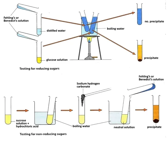
Benedict's or Fehling’s test: The food sample is dissolved in water, and a small amount of Benedict's reagent or Fehling’s reagent is added. The mixture is heated in a water bath for few minutes. A brick red precipitate signifies the presence of reducing sugars.
Test for Non-Reducing Sugar
The food sample is dissolved in water and heated with dilute hydrochloric acid to hydrolysis it into glucose and fructose. Dilute sodium hydroxide is added to neutralize the mixture until it stops fizzing. A positive results of the product with Benedict's or Fehling’s test indicates the presence of disaccharide non-reducing sugar (sucrose).
{nextPage}
Proteins
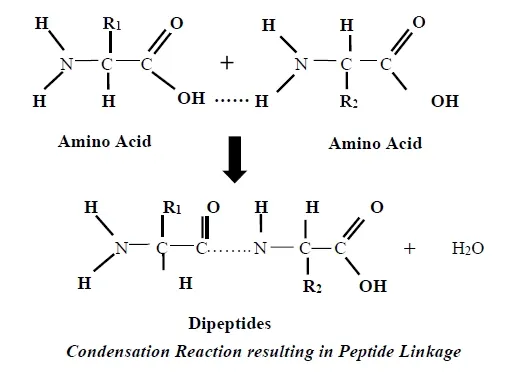
□ Non-essential amino acids are amino acids that the body can manufacture on its own.
□ Animal proteins such as meat, fish, eggs, milk contain all the
essential amino acids and they are known as first class protein.
□ Plant protein contains low amount of essential amino acids and they are described as second-class protein.
Sources of Proteins: meats, milk, fish, eggs, beans, nut butters, cowpea, crayfish and wheat.
Functions of Proteins
□ Formation of tendons and cartilage
□ Production of transport proteins such as hemoglobin and lipoproteins
□ Production of antibodies
□ Proteins are involved in muscle contraction and movement e.g. actin and myosin.
□ Repair body cells and make new ones.
Test for Proteins
□ Biuret test: An aqueous food sample is treated with an equal volume of dilute sodium or potassium hydroxide, followed by a few drops of 1% copper (II) sulphate. If the solution turns purple, protein is present.
Lipids
Lipids are large organic compounds containing carbon, hydrogen and oxygen. The proportion of oxygen is far smaller. Lipids includes: fats and oils, phospholipids and steroids.
□ Triglycerides are esters made up of 3 molecules of fatty acids and 1 molecule of glycerol.
□ Fatty acids are long chains of hydrocarbon making fats non-polar (insoluble in water).
□ Triglycerides that are solid at room temperature are classified as fats, and occur in animals. They contain saturated hydrocarbons. .
□ Triglycerides that are liquid at room temperature are called oils and originate in plants. Oils contain unsaturated hydrocarbon.
□ Unsaturated fats are derived from fatty containing double bonds within carbon chain. Unsaturated fats can be converted to saturated ones by the process of hydrogenation.
Source
of lipids
Functions of Lipids
□ Fats serve as a source of energy, containing about 37.8 KJ per gram of fat.
□ Protect vital organs such as heart, lungs and intestines.
□ Vitamins A, D, E, and K are fat-soluble, (meaning they can only be digested, absorbed, and transported in conjunction with fats).
□ Lipids also form the basis of steroid hormones.
Test for Lipids
□ Grease spot test/Brown paper test: Use a cotton swab to put samples of lipids on brown paper bag. Wait for few minutes to dry. Hold the paper up to a light source. The spot becomes translucent (allows light to pass through).
□ Sudan III (or Red) test: Add 2ml of any oil and 2ml of water to a test tube. Then add 2-5 drops of Sudan III to the mix. Shake. Sudan III will stain the fat molecules red.
Water
Water is a chemical compound with the chemical formula H2O.
A water molecule contains one oxygen and two hydrogen atoms connected by
covalent bonds.
□ It provides no calories (energy) or organic nutrients.
□ Water is a good polar solvent and is often referred to as the universal solvent.
Sources of water: water in drinks pineapple, coconut, water melon, water in plants
Functions of Water
□ Water forms the major component of the blood and lymph
□ Water also helps the blood carry oxygen and nutrients to all parts of the body.
□ It forms the major component of digestive juice.
□ Water helps formation of urine.
□ It helps removal of waste products.
Vitamins
Vitamin is an organic compound required by an organism as a vital
nutrient in limited amounts. The name is from vital and amine,
meaning amine of life. Vitamin cannot be synthesized by an organism, and must
be obtained from the diet.
Vitamins are classified as either water-soluble or fat-soluble.
Ø Fat-soluble vitamins: include A, D, E, and K. They are absorbed through the intestinal tract with the help of lipids (fats).
|
Vitamins |
Food Sources |
Functions |
Deficiency |
|
Vitamin A (Retinol) |
orange, leafy vegetables, carrots, egg-yolk, milk, fish |
healthy teeth, bones, soft tissue, and skin |
Night-blindness |
|
Vitamin B1 (Thiamine) |
pork, oatmeal, brown
rice, vegetables, potatoes, liver, eggs, palm wine |
Carbohydrate metabolism,
aid in cellular respiration, aid in synthesis of ribose |
Beriberi |
|
Vitamin B2 (Riboflavin) |
popcorn, milk, beans, green vegetables, mushroom, egg |
for growth; production of red blood cells |
Skin and corneal lesions |
|
Vitamin B3 (Niacin) |
Same as B2 |
maintain healthy skin and
nerves; has cholesterol-lowering effects |
Pellagra, Liver damage |
|
Vitamin C (Ascorbic acid) |
fruits and vegetables, liver |
maintain healthy tissue; promotes wound healing, |
Scurvy |
|
Vitamin D (Calciferol) |
fish, eggs, liver,
mushrooms, fortified cereals, fortified milk, cheese, yogurt, butter |
helps the body to absorb
calcium; maintenance of healthy teeth and bones |
Rickets |
|
Vitamin E (Tocopherols) |
fruits and vegetables, nuts, seeds, papaya, mango, oil |
formation of red blood cells, spermatogenesis, antioxidant |
Sterility in males, anaemia, miscarriages |
|
Vitamin K (phylloquinone) |
leafy vegetables, egg yolks,
liver, cereals, fish, beef |
important in blood
clotting |
Bleeding diathesis |
Minerals
Mineral nutrients are the chemical elements required by living organisms, other than carbon, hydrogen, and oxygen that are present in organic molecules.
|
Mineral |
Sources |
Functions |
Deficiency |
|
Potassium |
Legumes, potato skin, tomatoes, bananas, yams, soybeans |
heart and nerve health, is a systemic electrolyte |
muscle cramps |
|
Chlorine |
Table salt (sodium
chloride) |
production of HCl in the
stomach and maintenance of tissue fluid |
Mental apthy, muscle
cramps |
|
Sodium |
Table salt, vegetables, milk |
is essential in co-regulating ATP with potassium; transmission of
nerve impulses |
Mental apthy, muscle cramps |
|
Calcium |
Bones, eggs, fish, green
leafy vegetables, nuts |
is needed for muscle,
heart and digestive system health, builds bone, supports synthesis and
function of blood cells |
Rickets, stunted growth
and osteoporosis. |
|
Phosphorus |
Red meat, fish, poultry, bread, rice, oats |
required component of bones, essential for energy processing,
formation of nucleic acids |
Rickets |
|
Magnesium |
Nuts, soy beans, cocoa
mass, vegetables, fruit juice, beans, |
required for processing
ATP and transmission of nerve impulses |
Hypertension (high blood
pressure) |
|
Iron |
Red meat, leafy green vegetables, fish (tuna, salmon), eggs, dried
fruits, beans |
required for many enzymes, and for hemoglobin and some other proteins |
Anaemia |
|
Iodine |
Iodized salt, salt
fortified with iodine, sea fish |
required for the
synthesis of thyroxine |
Goitre, reduced growth |
|
Sulphur |
Eggs, meat, milk, fish, |
for synthesis of amino acids and many proteins (skin, hair, nails,
liver, and pancreas) |
No deficiency disease known |
BALANCED DIET
Balanced diet is a diet which contains all the essential food
nutrients in their correct proportion. Such diet must provide enough energy,
enough material for growth and replacement of worn-out tissues and must give
resistance against disease.
The constituents of balanced diet are roughly in the following proportions:
|
Carbohydrates |
about 50% |
|
Lipids |
25-30% |
|
Proteins |
10-25% |
|
Minerals and Vitamins |
a very small proportion but very necessary |
|
Water |
large amount |
{nextPage}
DENTITION
It is the type, number and arrangement of teeth in a given species of organism.
Type of Dentition
1. Homodont dentition: is when teeth are of the same size and shape. e.g. as in reptiles, amphibians, and fish.
2. Heterodont dentition: teeth that differ morphologically e.g. teeth in mammals.
Set of Teeth
o Deciduous Teeth or Milk Teeth: is a set of teeth found in infants, which fall out and replaced by permanent teeth. They are smaller and fewer in number.
o Permanent teeth: is a second set of larger teeth found in the adult that succeed the milk teeth.
Ø Monophyodont: is a dentition of animals with only one set of teeth throughout life.
Ø Diphyodont: is a dentition in animals that have two sets of teeth. E.g. humans. The first set (milk teeth) normally starts to appear at about six months of age. It has the same dental formula as the permanent teeth except the absence molars.
Ø Polyphyodont: is a dentition of animals in which the teeth continuously discard and replaced.
Types of Teeth
There are four distinct types of teeth in mammals:
Incisors
Canines
Premolars (also known as bicuspids)
Molars
· used for chewing or grinding food and for crushing or cracking bones
· the last molar teeth in each jaw to appear in humans are known as wisdom teeth
Enamel
Ø It forms a hard biting surface.
Dentine
o It decays more rapidly and subject to severe cavities if not properly treated
Pulp Cavity
Cement
Periodontal membrane
{nextPage}
Dentition of Herbivores
Herbivores are animals that feed on plant materials, e.g. sheep, goat, rat, rabbit, cow and donkey.
Adaptive Features of Herbivores
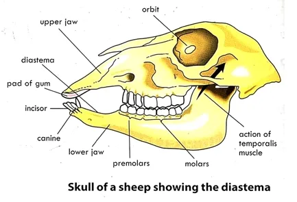
o presence of upper horny pad which allow the lower incisors to cut through vegetation
o cheek teeth are cusped for grinding and chewing grass
o side-to-side movement of the lower jaw enable chewing of food (as the teeth grind sideways, the enamel is worn out, exposing the dentine)
Dentition of Carnivores
Carnivore is an organism that feed on flesh and bones of animals often captured alive e.g. dog, lion, cat, tiger. Most carnivores are cannibals; they eat the flesh of their own kind.
Adaptive Features of Carnivores
o canines are large, long and pointed for piercing and holding prey
o premolars are sharp and pointed for tearing flesh
o the last upper premolar and first lower molar has modified to form the carnassial teeth for shearing meat.
o cusped premolars and molars for cracking bones
Dentition of Omnivores
Omnivores feed on both plants and animals e.g. human beings, apes, monkeys, bush pigs and bear. The teeth of omnivores have very little specialization. They have a combination of carnivore and herbivore teeth characteristics.
Adaptive features of omnivores
o cheek teeth have flat broad surface with cusps for grinding and crushing food.
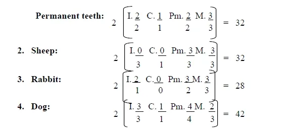
Dental Formula
Dental Care
o Regular brushing, at least twice a day to avoid formation of plaque.
o Use fluoride toothpaste or mouthwash to prevent dental decay.
o Avoid sugary foods.
o Visit dentist regularly.
o Eat food containing calcium, phosphorus, vitamin B, C, D for strong and healthy teeth.
o Avoid biting objects or nails to prevent cracking.
o Avoid using toothpicks which may lead to gum disease.
Dental Diseases
Dental diseases may affect the teeth, the gums, or other tissues of
the mouth. Dental diseases can affect the ability to chew, smile, or speak
properly and even lead to loss of tongue or teeth.
o Plaque: is a biofilm of saliva, mucus, bacteria food residues that form on the teeth. Plaque buildup, lead to tooth decays or periodontal problems.
Tooth Decay (Dental Caries)
Tooth decay is caused by acid-producing bacteria. They cause damage in the presence of sugar by producing lactic acid. The acid dissolves the calcium and phosphorus in the enamel. This process is known as "demineralisation" and it leads to tooth destruction.
Causes of Tooth Decay
· Lack of calcium in the diet
· Lack of vitamin D
· Failure to brush teeth after meal
Symptoms of Tooth Decay
o Uncomfortable when eating food, dinking hot or cold drink
Treatment of Tooth Decay
Periodontal Disease
This is the inflammation of the periodontal membrane and the cement as a result of bacterial infection. The bacteria eat the gum tissue, causing it to pull away from the teeth. This space between the tooth and gum, called periodontal pocket, can traps even more bacteria.
Causes of Periodontal Dises
o Lack of vitamin E in diet
Symptoms of Periodontal Disease
Treatment of Periodontal Disease
If the disease is not too severe it is possible to be treated with
fluoride rinse and fluoride toothpaste to remove the plaque, but once the
infection has progressed antibiotics will be needed to kill the bacteria.
You May like this Product: Check Out
{nextPage}
ENZYMES
Enzymes are biological catalysts which are protein in nature that speed up the rate of metabolic reaction in biological systems without themselves being altered in the process.
o Enzymes contain non-protein
components known as coenzymes, or cofactors which are essential for
their function. E.g. metal ions or water soluble vitamins.
o Enzyme (inactive) without its cofactor is known as the apo-enzyme and the active form i.e. enzyme plus cofactor is called the holo-enzyme.
Types of Enzymes
1. Exoenzyme: is an enzyme which is secreted by a cell and works outside the cell that produced it. It is usually used for breaking up large molecules that would not be able to enter the cell. E.g. Digestive enzymes and some clotting factors.
2. Endoenzyme: is an enzyme that functions within the cell that produced it was. E.g. dehydrogenases and decarboxylases.
Characteristics of Enzymes
o Enzymes are proteins in nature.
o They are substrates specific (e.g. lipase is for lipids).
o Enzymatic reactions are reversible.
o Enzyme activity may be decrease by inhibitors.
o Remain chemically unchanged at the end of a reaction.
o Required in small quantities.
o The rate of enzyme reactions is influenced by enzyme concentration.
o Some are inactive and requires co-enzymes to activate them.
o Activity is affected by temperature, pressure, pH and the concentration of substrate.
Factors Affecting Enzymes
How pH Affect Enzyme Reaction
Effect of Enzyme Concentration on Enzyme Reaction
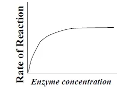
The rate of an enzyme reaction depends upon its own concentration. The rate of reaction is directly proportional to the enzyme concentration if the substrate concentration is fixed. As the amount of enzyme is increased, the rate of reaction increases. If there are more enzyme molecules than the amount needed, reaction rate increases as enzyme concentration increases but it levels off.Effect of Certain Molecules on Enzyme Reaction
Inhibitors and activators: Many molecules affect the rates of enzyme reactions. Some molecules bind to the enzyme or the substrate or the enzyme-substrate complex and lower the reaction rate. These are known as inhibitors. Similarly, some bind to the enzyme molecule and increase the reaction rate. These are known as activators.
"Lock and Key" Model
Enzymes are substrates specific. This is because both the enzyme and
the substrate possess specific geometric shapes that fit exactly into one
another. This is often referred to as the "lock and key"
model. An enzyme, as the key have
multi-dimensional shape, with pockets lined with specific amino acids. The
particular pockets that are active in catalyzing a reaction is called the
active sites of the enzyme. The active site is continuously reshaped by
interactions with the substrate. The active site is molded into the precise
position that enables the enzyme to perform its catalytic function.
{nextPage}
Human Digestive System: Functions, Processes, and Health Tips
It is a complex process that breaks down large organic molecules into smaller particles that the body can utilize as fuel.
It consists of alimentary canal and its accessory glands.
3. Deglutition: the swallowing of food.
4. Peristalsis: the rhythmic contraction and relaxation of the gust muscles which move food through the gastrointestinal tract or gut.
5. Digestion: is the mechanical and chemical breakdown of food into smaller components that are more easily absorbed into a blood stream. Digestion is a form of catabolism: a breakdown of large food molecules to smaller ones.
6. Absorption: the process whereby soluble end products of digestion pass through the intestinal walls into blood and lymph by osmosis, active transport and diffusion.
7. Assimilation: the process by which the absorbed food is used by the body cells and tissues.
Gastrointestinal Tract (Alimentary Canal)
Mouth (Oral Cavity)
Mouth is an organ for receiving
food and breaking up large organic compounds. In the mouth, food is digested
mechanically by biting and chewing. Function of the Tongue
o it allows the mixing of food with saliva
Oesophagus
Stomach
It is a small, 'J'-shaped pouch, which stores food temporally It is located on the left upper part of the abdominal cavity. It has two sphincters; the oesophageal or cardiac sphincter, found in the cardiac region and the pyloric sphincter dividing the stomach from the small intestine.The Role of Stomach in Digestion
o Production of gastric juice which contains pepsin for protein digestion.
Intestine
The intestine or bowel extends from the pyloric sphincter of the stomach to the anus. It consists of two segments, the small intestine and the large intestine.
The small intestine is further subdivided into the duodenum, jejunum and ileum.
The small intestine possesses numerous projections called villi (singular: villus).
Individual villus cells have microvilli which greatly increase absorptive surface area. Each villus contains blood vessels and a lacteal (lymph vessel).
Large intestine is subdivided into the cecum, colon and rectum.
The transverse section of the alimentary canal reveals;
o Mucosa: is the innermost layer of the
gut surrounding the lumen. It can be divided into the epithelium, lamina
propria,
and the muscularis mucosae. The epithelium and lamina
are filled with gastric glands that secrete mucus, hydrochloric acid, pepsin
and rennin. Muscularis mucosa is a thin layer of smooth muscle that contracts
and relaxes to move the food forward.
o Submucosa: consists of a dense irregular
layer of connective tissue with large blood vessels, lymphatics, and nerves.
o Adventitia or Serosa: consists of an
inner circular muscle layer and a longitudinal outer muscle layer. The circular
muscle layer prevents food from traveling backward and the longitudinal
layer shortens the tract.
The coordinated contraction of these layers is called peristalsis.
{nextPage}
Accessory Digestive Gland
An accessory digestive gland (or organ) is a gland that is not a part of the digestive tract. Accessory organs include the salivary glands, liver, gallbladder, and pancreas.
Salivary Glands
The three salivary glands (parotid, submandibular, and sublingual) secrete saliva, which contains enzymes that initiate breakdown of carbohydrates.
The Role of Saliva in Digestion
o Contains ptyalin (salivary amylase) which convert cooked starch into maltose
The Pancreas: Essential Functions and Roles in Digestion
The pancreas is a vital organ located in the abdomen, behind the stomach. It has both endocrine and exocrine functions, playing a crucial role in digestion and regulating blood sugar levels. The pancreas is a key player in the digestive system, contributing to both the chemical breakdown of food and the regulation of metabolic processes.
Exocrine Functions
1. Production of Digestive Enzymes
- Function: The pancreas produces digestive enzymes that are essential for breaking down carbohydrates, proteins, and fats in the small intestine.
- Enzymes: The main enzymes produced by the pancreas include:
- Amylase: Breaks down carbohydrates into simple sugars.
- Proteases (e.g., trypsin, chymotrypsin): Break down proteins into smaller peptides and amino acids.
- Lipase: Breaks down fats into fatty acids and glycerol.
2. Secretion of Bicarbonate
- Function: The pancreas also secretes bicarbonate into the small intestine. This alkaline solution neutralizes the acidic chyme (partially digested food) coming from the stomach, creating a suitable environment for the action of digestive enzymes.
Endocrine Functions
1. Regulation of Blood Sugar Levels
- Insulin Production: The pancreas produces insulin, a hormone that lowers blood glucose levels by facilitating the uptake of glucose into cells and promoting the storage of glucose as glycogen in the liver and muscles.
- Glucagon Production: The pancreas also produces glucagon, a hormone that raises blood glucose levels by stimulating the breakdown of glycogen into glucose in the liver.
2. Maintenance of Homeostasis
- Blood Sugar Balance: The balance between insulin and glucagon ensures stable blood sugar levels, which is essential for normal metabolic function and energy supply.
Disorders of the Pancreas
Several conditions can affect pancreatic function, including:
Diabetes Mellitus: A chronic condition characterized by high blood sugar levels due to insufficient insulin production or impaired insulin action.
- Type 1 Diabetes: An autoimmune condition where the body attacks insulin-producing cells in the pancreas.
- Type 2 Diabetes: A condition characterized by insulin resistance and eventually reduced insulin production.
Pancreatitis: Inflammation of the pancreas, which can be acute or chronic. It can result from conditions such as gallstones, chronic alcohol consumption, or certain medications.
Pancreatic Cancer: A malignant growth in the pancreas that can affect both endocrine and exocrine functions.
Importance of Pancreatic Health
Maintaining pancreatic health is crucial for:
- Effective Digestion: Proper enzyme production and secretion ensure efficient digestion and nutrient absorption.
- Blood Sugar Regulation: Healthy insulin and glucagon production is vital for maintaining stable blood sugar levels and preventing metabolic disorders like diabetes.
- Overall Well-being: Proper pancreatic function supports overall metabolic balance and energy regulation.
For more detailed information on the pancreas and its functions, visit these resources:
Liver
The liver is a reddish-brown organ with four lobes of unequal size
and shape. It is the largest internal organ (the skin being the largest organ)
and the largest gland in the human body. It is located in the right upper
quadrant of the abdominal cavity, resting just below the diaphragm. The liver
lies to the right of the stomach and overlies the gallbladder. It is connected
to two large blood vessels; one called the hepatic artery and other called the portal
vein. The hepatic artery carries blood from the aorta, whereas the
portal vein carries blood containing digested nutrients from the entire
gastrointestinal tract, spleen and pancreas to the liver.
Function of the Liver
The liver has a wide range of functions, including;
o Formation of red blood cells in a fetus.
o Production of bile
o The liver breaks down or modifies toxic substances in a process called detoxification.
o The liver converts excess amino acids into urea in a process known as deamination.
o The liver produces heat for body temperature regulation.
o Decomposition of red blood cells.
o Storage of a glycogen, vitamin and iron.
o Storage of blood.
o The liver produces albumin, the major component of blood serum.
o It breaks down insulin and other hormones
o The liver regulates blood sugar level by converting excess glucose into glycogen for storage and converts glycogen back to glucose when the level of blood sugar falls.
o It synthesized organic compound such as glucose, cholesterol and lipoproteins.
o It produces coagulation factors such as fibrinogen, prothrombin, and antithrombin.
Disorders of the Liver
Several conditions can affect liver function, including:
- Hepatitis: Inflammation of the liver, often caused by viral infections or excessive alcohol consumption.
- Cirrhosis: Scarring of the liver tissue due to long-term damage, which can impair liver function.
- Fatty Liver Disease: Accumulation of fat in liver cells, often associated with obesity and metabolic disorders.
- Liver Cancer: A malignant growth in the liver that can develop due to chronic liver disease or hepatitis.
Importance of Liver Health
Maintaining liver health is crucial for overall well-being. A healthy liver ensures efficient digestion, detoxification, and nutrient processing. Lifestyle choices such as a balanced diet, regular exercise, and moderate alcohol consumption can help support liver function and prevent liver diseases.
For more detailed information on the liver and its functions, visit these resources:
- Mayo Clinic: Liver Functions
- National Institute of Diabetes and Digestive and Kidney Diseases (NIDDK): Liver
Digestion in the Mouth (Oral Cavity)
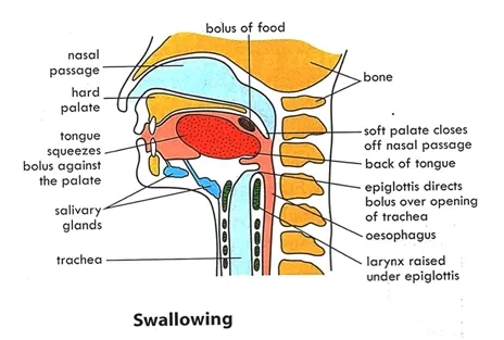
Digestion begins in the Mouth, where food is chewed or masticated with the teeth. Saliva is secreted by three pairs of salivary glands in the oral cavity, and is mixed with the chewed food by the tongue. Saliva moistens the food. It contains digestive enzymes, salivary amylase, which breakdown starch into maltose. It also contains mucus that soften the food and form it into a bolus. An additional enzyme, lingual lipase, hydrolyzes fats/oil into partial glycerides and fatty acids. The tongue manipulates the food and roll it into a bolus which is swallowed into the stomach. The bolus moves by peristalsis through the oesophagus.
For more detailed information on the role of the mouth in digestion, visit these resources:
- Mayo Clinic: Digestion and the Digestive System
- National Institute of Diabetes and Digestive and Kidney Diseases (NIDDK): Digestive System
Digestion in the Stomach
The bolus enters the stomach through the esophageal sphincter. The
presence of food in the stomach stimulates the stomach walls to secrete a
hormone called gastrin. Gastrin stimulates the gastric glands in the stomach
to secrete gastric juice. The
gastric juices contain hydrochloric acid, mucus and proteases (pepsinogen
and prorennin).
o Rennin digests milk protein, caesinogen into caesin (curds) i.e. curdling the milk.
o Pepsin breaks down proteins into peptides.
Functions of Hydrochloric Acid
The food is partially digested and semi-liquid, referred to as chyme. Chyme slowly passes through the pyloric sphincter and into the duodenum of the small intestine.
Digestion in the Small Intestine
In the duodenum, acidic chyme from stomach triggers the cells of the duodenum to produce secretin. Secretin stimulates the pancreas to produce pancreatic juice. It also stimulates the liver to secrete bile. The presence of food in the duodenum also stimulates the release of another hormone called cholecystokinin, which function the same way as the secretin.
Pancreatic Juice
o Trypsin and Chymotrypsin digest proteins to peptides.
o Pancreatic lipase digests fats to glycerol and fatty acid.
Bile
Bile is a greenish-yellow fluid that contains water, salts and bile pigment (bilirubin, biliverdin), but has no enzyme. It is produced by the liver when worn out red blood cells are broken down. It is then stored in the gallbladder where it released via the bile duct.
Functions of Bile
o Eliminates waste products such as bilin and bile acids from the blood.
Intestinal juice
The Table Below Shows Digestive Enzymes Grouped by Source of the Enzyme
|
Source |
Enzyme |
Food (Substrates) |
Product |
|
Mouth (salivary glands) |
Salivary amylase |
Polysaccharides |
Maltose |
|
Stomach |
Pepsin Rennin |
Proteins Milk protein
(caseinogens) |
Peptides Caseins |
|
Pancreas |
Pancreatic amylase Trypsin & chemotrypsin Lipase |
Polysaccharides Proteins Fats |
Maltose Peptides Fatty acid and glycerol |
|
Small Intestine |
Maltase Peptidases Sucrase Lactase Lipase Erepsin Enterokinase Nucleotidase |
Maltose Peptides Sucrose Lactose Fats Peptides Trypsinogen Nucleotides |
Glucose Amino acids Glucose and fructose Glucose and galactose Fatty acid and glycerol Amino acids Trypsin Sugar, phosphorus and nitrogen base |
Large Intestine
The large intestine absorbs water from the chyme. Food products that cannot go through the villi form feces. Faeces contain cellulose (dietary fiber), water, mucus, dead cells, bile pigments and bacteria. Bacteria of large intestine metabolize bilirubin to urobilinogen, which is responsible for the brown color of feces.
{nextPage}
Absorption
Most absorption in the small intestine occurs in the duodenum.
Adaptation of Small Intestine for Absorption of Nutrients
□ Thin epithelial cells of the villi (one cell thick), allow easy diffusion of nutrients.
□ Epithelial cells secrete mucus to prevent friction
□ Epithelial cells contain numerous mitochondria which provides energy/ATP for active transport
□ Lacteal (lymphatic vessels) to transport fatty acids and glycerol.

Absorption of fatty acids and glycerol
Fatty acid and glycerol enter the villi by diffusion. In the villi, they reassembled into fats (triglycerides) and package into chylomicrons or liposomes. They move into the lacteals for transport to the rest of the body through the lymphatic system.
Absorption of Glucose and Amino acids
Diffusion and active transport is responsible for the movement of glucose and amino acids. Villus capillaries collect amino acids and glucose taken up by the villi into the blood stream. Blood containing the nutrients is transported via the hepatic portal vein to the liver for filtering, removal of toxins, and nutrient processing.
Assimilation
Assimilation is the process by which the absorbed
food is used by the cells and tissues of the body i.e., the metabolism of
end-products of digestion.
Glucose metabolism
Glucose (end-product of carbohydrates) is the only sugar used by the
body cells to provide energy. Therefore, all monosaccharides must be converted
into glucose metabolite by liver. In the liver, insulin produced
by pancreatic cells convert excess glucose to glycogen for
storage in the muscle or liver. This reduces the level of glucose in the blood.
In contrast, glucagon is secreted into the
bloodstream by pancreatic cells. It promotes the breakdown of glycogen in the
liver to release glucose into the blood. Glucagon also promotes gluconeogenesis,
a process involving the synthesis of glucose from amino acids or fatty acids.
Amino acids metabolism
Ingested proteins are broken down into free amino acids by enzymes
called proteases. Amino acids are used for protein
biosynthesis. Excess amino acids are converted to glucose through gluconeogenesis if
glucose run short. Excess amino acids cannot be stored, and can be poisonous if
accumulated in the body. They are converted to a less toxic compound
called urea by the liver. This process is referred to as deamination,
which involved the removal of the amine group from amino acids. The remaining
organic acids or keto-acids are converted into glycogen for storage.
Fatty Acids and Glycerol Metabolism
Fatty acids and glycerol recombine, after absorption to form fats in the
body. They are oxidized to release energy. The liver also converts excess
carbohydrates and proteins into fats which are stored in the adipose tissues.
Excess fats are stored around vital organs such as heart, digestive,
reproductive and excretory organs.
Roughage (Dietary fibre or Bulk)
Roughages are the indigestible part of plants consumed as food. Roughage
consists of primarily of cellulose, a component of plant cell walls. It found
in fruits, vegetables and grains.
Functions of Roughage
Malnutrition
Malnutrition is a diet condition caused by a
deficiency or excess of one or more essential nutrients in the diet. The intake
of inadequate nutrients results in deficiency diseases. Some deficiency
diseases are
Marasmus
Marasmus is a form of severe protein deficiency and is one of the forms of
protein-energy malfunction. A child with marasmus looks emaciated. Body weight
may be reduced to less than 80% of the average weight.
Signs and symptoms of Marasmus
o extensive tissue and muscle wasting
o dry, cracked skin
Treatment
It can progress to the point of no return when the body's machinery for
protein synthesis degrades.
Kwashiorkor
Kwashiorkor is an acute form of childhood protein-energy malnutrition. Insufficient protein consumption, but with sufficient calorie intake, distinguishes it from marasmus. It can have a long-term impact on a child's physical and mental development, and may lead also to death.
Signs and symptoms of Kwashiorkor
Treatment of Kwashiorkor
It can be
treated by adding protein to the diet.
Constipation
Constipation also known as costiveness refers to
the difficulty in emptying the bowel. It is a common cause of painful defecation.
Severe constipation includes obstipation (failure to
pass stools or gas) and fecal impaction, which
can become life-threatening. Constipation is a symptom, not a disease,
therefore effective treatment requires first determining the cause.
Causes of Constipation
Symptoms of Constipation
Prevention of Constipation
Indigestion
Indigestion also known as
dyspepsia is a medical condition characterized by recurrent pain in the upper
abdomen, upper abdominal fullness and feeling full earlier than expected when
eating.
Causes of Indigestion
Signs and Symptoms pf Indigestion
Ways of preventing indigestion
Treatment of indigestion: by medication or drug therapy



.webp)

.webp)


