CELL CYCLE AND CELL DIVISION
Objectives
This blog post provides readers with the following objectives. The reader will be able to:
o Explain some terms under cell cycle
o Outline the process of the cell cycle
o Describe the process mitosis and meiosis.
{getToc} $title={Table of Contents} $count={Boolean} $expanded={Boolean}
CELL CYCLE
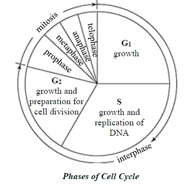
The cell cycle is the sequence of events that takes place in cells.
It leads to cell division and replication. In eukaryotes, the cell cycle can be
divided into four distinct phases: G1
phase, S phase (synthesis), G2 phase and M phase. The G1, S phase and
G2 phase together are known as interphase. The M phase or the
mitotic phase (cell division) occurs
after interphase. Cell division consists of two phases: nuclear division followed
by cytokinesis. Nuclear
division divides the genetic material in the nucleus, while cytokinesis divides
the cytoplasm.
There are two kinds of nuclear division: mitosis and meiosis.
INTERPHASE
It is the resting stage or the non dividing phase of the cell cycle. It takes about 90% of the cell's life span.
The cell prepares itself, grows and accumulates nutrients for DNA replication and division.
The nucleus is visible and the chromosomes are uncoiled and invisible. The cells grow by producing proteins and cytoplasmic organelles.
The Stages of Interphase
Interphase consists of three main stages:
G1 Phase (Gap 1): During the G1 phase, the cell grows and synthesizes proteins necessary for DNA replication. This stage is crucial for cell function and prepares the cell for the next phase. Learn more about the G1 Phase in Cell Cycle from Khan Academy.
S Phase (Synthesis): In the S phase, DNA replication occurs. The cell’s genetic material is duplicated, ensuring that each daughter cell will receive an identical set of chromosomes. This process is fundamental for genetic continuity. For a detailed explanation of DNA replication, visit DNA Replication on National Institutes of Health (NIH) (.gov).
G2 Phase (Gap 2): The G2 phase involves further cell growth and the production of proteins needed for mitosis. During this phase, the cell checks for any DNA errors and makes necessary repairs. This ensures that the cell is ready for the mitotic phase. Explore the G2 Phase and its Functions on Frontiers in Cell and Developmental Biology for more insights.
The Importance of Interphase
Interphase is crucial for several reasons:
- Cell Growth: The cell increases in size and synthesizes essential proteins, enabling it to function properly and support division.
- DNA Replication: Accurate duplication of the genetic material is vital for maintaining genetic integrity and preventing mutations.
- Preparation for Division: Interphase ensures that the cell has all the necessary components to successfully undergo mitosis or meiosis, leading to the production of daughter cells.
Disorders Related to Interphase
Errors during interphase can lead to various disorders, including cancer. Abnormalities in DNA replication and repair mechanisms can result in uncontrolled cell growth. For an overview of cancer related to cell cycle dysregulation, visit Cancer and the Cell Cycle on the National Cancer Institute’s website.
MITOSIS
Mitosis is a type of cell division in which a single cell produces
two genetically identical daughter cells. The daughter cells contain the same
genetic material as the parent cell. It is the way in which new body cells are
produced for growth and repair.
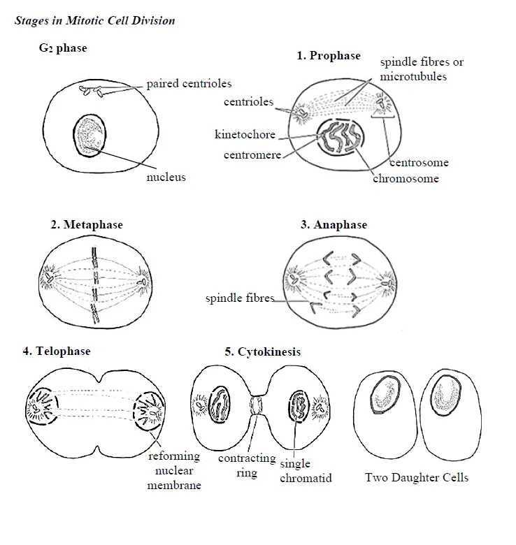
There are four stages of mitosis: Prophase, Metaphase, Anaphase, Telophase.
Prophase
1. The chromosomes condense and become visible.
2. Each chromosome is seen as two chromatids joined together at a point called centromere.
3. The nucleolus disappears and nuclear membrane breaks down.
4. Centrioles move to opposite ends of the cell.
5. Spindle fibers extend from the centrioles.
Metaphase
1. Chromosomes arranged at the equator of the cell.
2 Spindle fibers attach to kinetochores of chromosomes.
Small disc-shaped structure at the surface of the centromere is called kinetochore
Anaphase
1. Spindles begin to shorten and separate the sister chromatids at the centromere.
2. Spindle fibers continue to shorten, pulling the chromatids to opposite poles. This ensures that each daughter cell gets identical sets of chromosomes
Telophase
1. Chromatids reach the poles.
2. The nuclear membrane and nucleolus reappears.
3. The chromosomes uncoil.
4. The spindle apparatus breaks down.
5. Cytokinesis may also begin during this stage
Cytokinesis
Cytokinesis is not a phase of mitosis. It is a separate process necessary for completing cell division.
In animal cells a cleavage furrow containing a contractile ring develops at the equatorial plane separating the nuclei.
In plant cells, a cell plate is formed across the middle of the cell to divide the cytoplasm and cell wall is between the two daughter cells.
Significance of Mitosis
1. It brings about growth.
2. The daughter cells have the same genetic material as that of the parent cell.
3. It is responsible for development of a single-celled zygote into a multicellular organism.
4. The daughter cells have the same characters as those of the parent cell.
5. It helps in repairing and replacing dead or damaged cells.
6. It’s a means of reproduction in unicellular organisms.
7. It may result in uncontrolled growth of cells leading to cancer or tumor.
Difference between Mitosis in Plant and Animal Cells
|
Plant cell |
Animal cell |
|
Centriole is absent |
Centriole present |
|
Does
not form aster |
Aster is formed |
|
Cell plate is formed to divide daughter
cell |
Does not form cell plate |
|
Occurs
in meristem |
Occurs in all cells |
MEIOSIS
Meiosis is a reduction division of a diploid cell to form haploid daughter cells. Haploid cells contain half the number of chromosomes as the parent cell. It takes place in all sexually reproducing organisms. Meiosis is restricted to the gonads or sex cells. Meiosis is divided into meiosis I and meiosis II.
Meiosis I
It involves the splitting of parent cell and separation of the homologous chromosomes from each other into different cells.
The resulting daughter cells contain one entire haploid set of chromosomes.
It produces two haploid cells (N chromosomes, 23 in humans).
Prophase I
1. Prophase I is the longest phase of meiosis I.
2. The chromosomes condense and become visible within the nucleus
3. The nuclear membrane begins to disintegrate.
4. The centrioles form and move toward the poles.
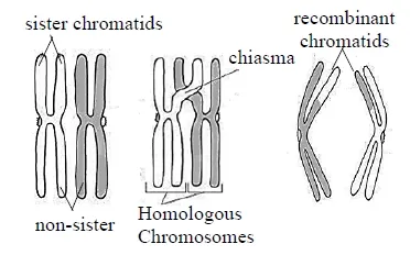
5. In this phase, there is exchange of DNA between homologous chromosomes, this process is known as homologous recombination.
6. The homologous chromosomes pair up forming bivalents or tetrads. Each chromosome comes from each parent.
7. Pairing of homologous chromosomes is called synapsis. At the stage of synapsis, the non-sister chromatids may cross-over at points called chiasmata. Crossing over result in exchange of fragments.
8. Crossing over serves to increase genetic diversity by creating four unique chromatids
Metaphase I
1. Microtubules or spindle fibers grow from the centrioles.
2. The bivalents line up along the cell equator and attach to spindle fibers at the centromeres.
Anaphase I
The homologous chromosomes separate and move towards the opposite pole of the cell while sister chromatids remain associated at their centromeres.
Telophase I
2. The spindle fibers disappear.
3. The chromosomes uncoil and return back to the chromatin stage.
4. The process of cytokinesis occurs, creating two haploid daughter cells.
5. The daughter cells have half the number of chromosomes.
Meiosis II
Meiosis II is an equational division similar to mitosis, where the sister chromatids split forming 4 haploid cells, two from each daughter cells of meiosis I.
Prophase II
1. The nucleoli and the nuclear envelope disappear.
2. Chromosomes condense.
3. The centrioles move toward the poles.
Metaphase II
1. The spindle fibres grow from the centrioles and attach to the centromeres.
2. The sister chromatids line up along the cell equator.
3. The equatorial plane formed here is rotated by 90 degrees, compared to meiosis I and is perpendicular to the previous equatorial plane.
Anaphase II
1. The centromeres break and sister chromatids separate.
2. The sister chromosomes move towards the opposing poles.
Telophase II
1. Chromatids reach the opposite poles of the cells.
2. The nuclear membrane and nucleolus reappears.
3. The chromosomes uncoil.
4. The spindle apparatus breaks down.
5. Cleavage or cell wall forms which eventually produces a total of four daughter cells, each cell having haploid set of chromosomes.
Significance of meiosis
1. It ensures continuity of species and brings about variation.
2. It enables sexual recombination to occur by reducing the chromosome number by half.
Stages in Mitotic
Cell Division
Stages in Meiotic Cell Division
Difference between Mitosis and Meiosis
|
Mitosis |
Meiosis |
|
Nucleus divides only once |
Nucleus divides twice |
|
Chromosome number is maintained |
Chromosome number is halved |
|
No pairing of chromosomes |
Pairing occurs |
|
Chiasmata
are never formed |
Chiasmata are formed |
|
Crossing - over never occurs/ no exchange
of genetic materials |
Crossing - over occurs /exchange of genetic
materials occurs |
|
Daughter cells identical to parent cell |
Daughter cells generally different from parent cell |
|
Two daughter cells are formed |
Four daughter cells are formed |
|
Chromosomes
form single row at the equator |
Chromosomes form double
row at the equator |
|
Occurs in somatic cell |
Occurs in reproductive cell |
|
Occurs
in asexual reproduction |
Occurs in sexual
reproduction |
Get Your Free PDF!
Download our comprehensive guide to
CELL CYCLE AND CELL DIVISION
to boost your knowledge.
Download PDFClick the button above to get instant access to the PDF.


