THE NERVOUS SYSTEM AND ENDOCRINE SYSTEM IN HUMAN
This blog post provides readers with the following objectives. The reader will be able to:
Co-ordination is the ability to receive stimuli
and to respond appropriately to ensure the maintenance of a steady state and
survival of the organism. The two main system that bring about co-ordination are Nervous system and Endocrine system
Nervous System
The nervous system is divided into two principal parts.
1. Central nervous system (CNS) consists of the brain and spinal cord.
2. Peripheral nervous system (PNS) consists mainly of nerves (i.e., peripheral nerves), which connect the CNS to other parts of the body. The spinal nerves carry impulses to and from the spinal cord and cranial nerves carry impulse to and from the brain. The PNS is divided into sensory (afferent) and motor (efferent) divisions.
a. A sensory receptor is the specialized region of the body (internal and external) responsible for detecting a stimulus and transmit impulse to the CNS. Sensory receptors usually associated together to form sense or receptor organ. Examples include eye, ear, skin, nose and tongue.
b. Sensory (afferent) nerves transmit impulses from the receptors to the CNS. Sensory nerves which transmit impulses from the skin, skeletal muscles and joints are called somatic afferent, where as those which transmit impulses from the visceral organs are called visceral afferent.
c. Effector is a structure or an organ which responds to a stimulus e.g., muscles, glands.
d. Motor (efferent) nerves transmit impulses from the CNS to effectors organs, the muscles and the glands. The impulses activate muscles to contract and glands to secrete substance. The motor division in turn, has two subdivisions; the somatic nervous system and autonomic nervous system.
Nerve Cell or Neuron
Nerve cell is the structural and functional unit of the nervous system. Nerve cells are highly specialized cells that conduct nerve impulses from one part of the body to another.
Typical neuron consists of a cell body, and cell extensions or fibers called Dendrites and Axons.
Cell body: This is the enlarged portion of the neuron. It contains a well-defined nucleus and nucleolus that serves as the “biosynthetic center”, where macromolecules are produced. They have no centrioles and this makes them amitotic. Cell bodies within the CNS are frequently clustered into groups called nuclei whereas cluster of cell bodies in the PNS are referred to as ganglia.
Dendrites: These are thin, branched cytoplasmic extensions of the cell body. Dendrites receive signals from other neurons and transmit them on to the cell body.
Axons: The axon is the projection that conducts impulses away from the cell body. Some axons are surrounded by a myelin sheath. These are known as myelinated axons. Other axons do not have a myelin sheath and are called unmyelinated axons. Areas of the CNS that contain a high concentration of myelinated axons form the white matter. The gray matter of the CNS is composed of high concentrations of the cell bodies and dendrites which lack myelin sheath
Support Cells (glial or neuroglia)
Glial cells are
smaller, non-excitable cells that surround delicate neurons. The glial
cells include microglia, oligodendrocytes, and Schwann
cells.
1. Microglias are special type of macrophage that helps protect the CNS by engulfing invading microorganisms and dead neural tissue.
2. Oligodendrocytes wrap tightly around the nerve fibers, producing insulating covering called myelin sheaths.
3. Schwann cells, form myelin sheaths around the larger nerve fibers in the PNS, are functionally similar to oligodrocytes. Schwann cells also act as phagocytes to rid of a damaged nerve.
Functions of glial cells
Classification of Neurons
2. Motor (efferent) neurons conduct impulses out of the CNS to the effector organs (muscles and glands).
3. Intermediate or Relay are located entirely within the CNS. They connect the sensory and motor neurons with each other.
Generation and Transmission of Nerve Impulse
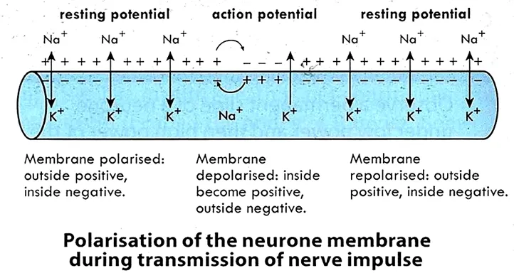
When axon is not conducting an impulse, the inside is negatively charged whilst outside is positively charged. This is called resting potential and the neuron is said to be polarized. The concentration of sodium ions (Na+) is greater outside the axon than inside and the concentration of potassium (K+) is greater inside the axon than outside. This difference in concentration is due to the action of the sodium-potassium pump, a membrane protein that actively transports Na+ out of and K+ into the axon across the membrane.
When stimulated, positive charges (Na+) flow into the
cell causing depolarization. If the stimulus causes the nerve membrane to
depolarize to certain level called threshold, an action potential occurs. At threshold, depolarization causes the sodium-gates to open allowing Na+
to diffuse into the cell. The potassium
gates open and allow K+ flows from the inside to the outside of
the cell. These changes in Na and K diffusion and the resulting changes in the
membrane potential constitute an action potential or nerve
impulse.
The action potential is an all-or-none phenomenon; it either happens completely or it does not happen at all. When depolarization is below a threshold value action potentials are not produced.
During the time that a patch of axon membrane is producing an action potential, it is incapable of responding to further stimulation. This is called the refractory period. As the neuron returns to its original resting state, the action of the Na pump expels the Na from the cell in exchange for K; this is referred to as repolarization.
Synaptic Transmission
Synapse is a connection between two neurons or between a
neuron and effector cell, located in the muscle or gland. The two nerve cells do not actually
touch; there is a microscopic space between them. This space is the synaptic
cleft or Synapse, and it occurs between the axon of
one neuron and the dendrites or cell body on the next neuron. The neuron that is carrying the
impulse to the synaptic cleft is called presynaptic neuron and one that
carries the impulse away is the postsynaptic neuron.
The axon of the presynaptic neuron terminates in small swellings
called synaptic knobs, which are filled with chemicals known as neurotransmitters.
Neurotransmitters are stored in small vesicles called synaptic vesicles. They are released in response to the action potential or impulse and diffuse across the synaptic cleft, to activate or stimulate the postsynaptic cell. That is the impulse is changed from electrical impulse to a chemical impulse (Electrochemical Impulses).
Substances that have been identified at neurotransmitters include acetylcholine,
norepinephrine,
serotonin,
dopamine,
histamine
and glycine.
THE BRAIN
The brain is an organ that serves as the center of the nervous system. The brain is protected by a bony structure called the cranium, membranes called the meninges, and a watery cushion called the cerebrospinal fluid. The brain consists of three major regions:
1. Forebrain (which includes Cerebrum, hypothalamus and pituitary glands)
2. Midbrain or Brain Stem
3. Hindbrain (Cerebellum, pons and medulla oblongata)
Cerebrum: is the largest and most obvious portion of the brain. The cerebrum consists of right and left hemisphere, which are incompletely separated by a longitudinal cerebral fissure. Each hemisphere of the cerebrum is divided into lobes or parts which are separated by sulci (singular: sulcus). The cerebrum consists of two layers. The surface layer, the cerebral cortex, is highly convoluted and composed of gray matter. Beneath the cerebral cortex, is the thick white matter of the cerebrum, which constitutes the second layer. The cerebral cortex controls all voluntary motor function. The cerebrum is concerned with intelligence, memory, will power, imagination, reasoning, vision, speech, smell and taste.
Hypothalamus: is small portion located below the thalamus. It performs numerous functions vital to body homeostasis. It regulates movement of food through the gut, body temperature, blood pressure and water balance. It controls hunger or thirst, anger, sleep, sex drive, fear and pleasure. The hypothalamus also controls the pituitary gland by producing hormones.
Midbrain: connects the forebrain to the hindbrain and the spinal cord. It is involved in functions such as vision, hearing, eye movement, and body movement.
Pon: is a rounded bulge on the outside of the brain, between the midbrain and the medulla oblongata. It is involved in motor control and sensory analysis. Some structures within the pons are linked to the cerebellum, thus are involved in movement and posture.
Medulla Oblongata: extends from the Pons and ends at the spinal cord. Externally, it resembles the spinal cord. It carries signals between the spinal cord and the brain. It regulates most involuntary and reflex action of the body such as rhythm of breathing, heartbeat, swallowing, vomiting, coughing and sneezing.
Cerebellum: is the second largest structure of the brain. It is situated behind
the pons. The cerebellum has a thin outer layer (cerebellar cortex) of gray matter and a thick deeper layer of white
matter. The cerebellum coordinates skeletal muscle contractions, posture and
balance.
THE SPINAL CORD
The spinal cord is a long, fragile tube-like structure that extends from
the base of the brain down to the bottom of the spine (spinal column). The
spinal cord is covered by three layers of tissue called meninges. The spinal cord and
meninges are contained in the spinal canal, which runs through the center of
the spine. The spine is made up of individual bones called vertebrae which protect the spinal cord.
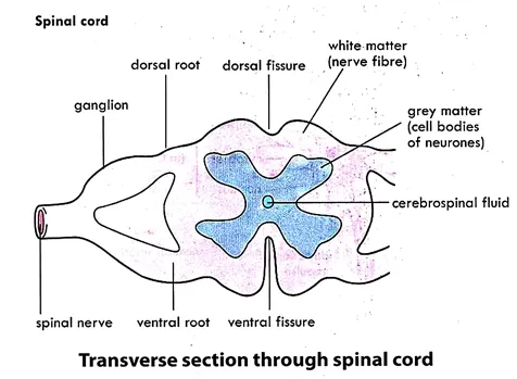
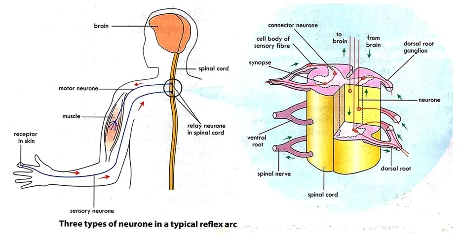
The spinal cord consists of white matter on the outside and grey matter in the shape of an H or butterfly on the inside. The grey matter contains dendrites and cell bodies. The front side of the spinal cord or ventral root contains motor nerves, which transmit information from the brain or spinal cord to muscles. The back or dorsal root contains sensory nerves, which transmit sensory information from other parts of the body through the spinal cord to the brain. There are a total of 31 pairs of spinal nerves. These carry impulses to and from the spinal cord.
Reflex Action and Reflex Arcs
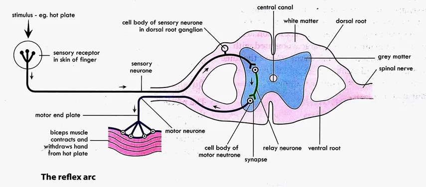
Reflex action is a fast, involuntary reaction or response to internal or external stimulus. Examples of reflex actions are breathing, eye blinking, iris size, and many protective actions such as moving away from a burning flame. It is a protective response to a situation in an attempt to maintain body homeostasis.
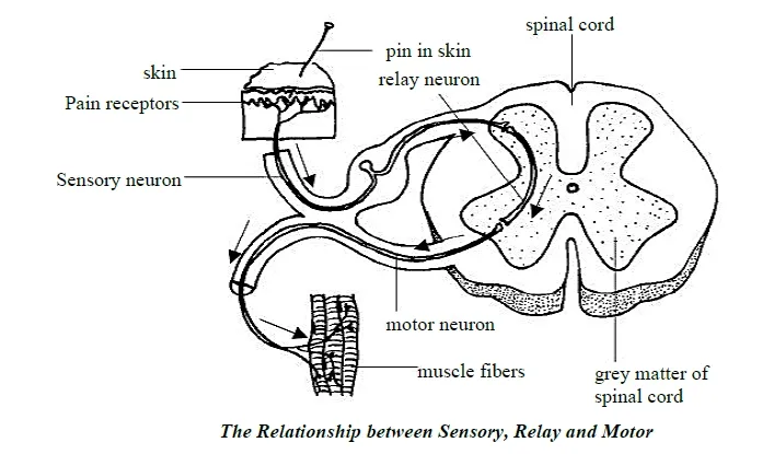
Reflex arc is the neurons that form the pathway along which an impulse travel during reflex action. The reflex arc has five basic components: sensory receptor, sensory neuron, an interneuron, motor neuron and an effector organ.
1. A receptor: the end of dendrite or some specialized receptor cell, as in special sense organ, that detect stimuli
2. A sensory neuron: a cell that conducts the impulses to the CNS.
3. An interneuron: a cell or cells within the CNS. These neurons may carry impulses to and from the brain or may take impulses across the spinal cord to the motor neurons.
4. The motor neurons: conducts impulse from the CNS to the effector (muscle).
5. An effector: a muscle or a gland outside the CNS that carries out response.
Types of reflexes
1. Monosynaptic reflexes involve only one CNS synapse e.g., Knee jerk
2. Polysynaptic reflexes involve two or more CNS synapses e.g. Pain withdrawal or pupil reflex
Importance of Reflex Action
1. They are automatic and fast, and occur without having had to be thought about
2. It serves as defense mechanism of the body against danger
3. Permit rapid response to stimuli
4. It helps organisms to anticipate certain events and initiate
Mechanism of Reflex Action
When we move our finger away from a flame, we are performing a withdrawal reflex. The stages of this reflex are as follows:
1. The finger is the receptor. It contains sensory neurons. Sensory neurons carry the impulse from the receptor to the spinal cord through the dorsal root.
2. An interneuron receives impulses from the sensory neuron and carries them across the spinal cord to the motor neurons in the ventral root. At the same time, another neuron takes the impulse to the brain.
3. The motor neurons take the impulse to the effector (muscle) and the finger is pulled away. At the same time, the impulse reaches the brain and we are aware of the pain.
A reflex arc that passes through the brain, is termed a cranial
reflex arc and the action involved is a cranial reflex action. Spinal
reflex arcs, however, pass through the spinal cord, producing spinal
reflex actions.
Somatic Nervous System
1. Somatic nervous system (SNS) is composed of nerve fibers that conduct impulses from the CNS to skeletal muscles. It is often referred to as the voluntary nervous system.
2. Voluntary actions: are action or activities of the body initiated under the direct control of the brain and therefore can be suppressed. It allows an individual to consciously control his/her skeletal muscles. E.g., walking, speaking, writing.
Autonomic Nervous System
1. Autonomic Nervous System (ANS) consists of motor nerve fibers that regulate the activities of smooth muscle, cardiac muscle and the glands. The autonomic nervous system controls internal body functions that are not under conscious control. ANS is referred to as the involuntary nervous system.
2. Involuntary actions: are actions or activities of the body that occur unconsciously hence not under control of the will and cannot be suppressed. E.g., include breathing, swallowing, sexual arousal, salivation, urination and pumping of the heart.
ANS has two functional subdivisions, the sympathetic and parasympathetic, which typically bring about opposite effects on the activity of the same visceral organs.
Sympathetic Nervous System: This part of the autonomous nervous system stimulates the activity of an organ as per the need. It speeds things up and prepares the body for activity. For example; during running, there is an increased demand for oxygen by the body. This is fulfilled by an increased breathing rate and increased heart rate.
Parasympathetic Nervous System: This part of the autonomous nervous system slows the down the activity of an organ and thus has a calming effect. During sleep, the breathing rate and the heart rate slow down.
Examples of sympathetic and parasympathetic system action on various organs
|
Effector |
Sympathetic |
Parasympathetic |
|
Eye |
Dilates pupil |
Constricts pupil |
|
Heart |
Increases rate of heart
beat |
Decreases rate of heart
beat |
|
Blood Vessels |
Constricts |
Dilates |
|
Sweat Glands |
Activates sweat secretion |
Inhibits sweat secretion |
|
Digestive tract |
Inhibits peristalsis |
Stimulates peristalsis |
|
Adrenal glands |
Stimulates
secretion |
Inhibits secretion |
|
Penis |
Promotes ejaculation |
Stimulates erection |
|
Salivary glands |
Inhibits secretion of saliva |
Stimulates secretion of saliva |
Difference between Voluntary and Involuntary Action
|
Voluntary Action |
Involuntary Action |
|
Controlled by the brain |
Controlled by the spinal cord |
|
Response
can delay |
Response is immediate |
|
Conscious response to stimulus |
Unconscious response to stimulus |
|
Path of
the nerve impulse is much longer |
Path of the nerve impulse
is by the shortest route |
|
Response is in skeletal muscle only |
Response is in skeletal or an internal
involuntary muscle |
{nextPage}
STRUCTURE OF MAMMALIAN EYE
The eye is the
organ for sight. It consists of spherical eyeball situated in a cavity in the skull called orbit. It is attached to the wall of
the orbit by six muscles called rectus muscles.
The wall of the
eyeball is composed of three layers; the outer sclera, the middle choroid and
the inner retina.
Sclera: is a white connective layer, which supports and maintains the shape of the eye ball. At the front of the eye the sclera is visible as the “white”.
Cornea: continuous with sclera but transparent, it allows light to enter the eye.
Choroid: lies beneath the sclera. It contains many blood vessels, and nourishes the cell. Its inner surface is highly pigmented and absorbs light ray
Iris: is a pigmented disc with two sets of muscles. It is the colored part of the eye that controls the amount of light that enters the eye through pupil.
Pupil: is a circular opening in the center of the iris.
Lens: a transparent disc held in place by suspensory ligament. It focuses images of objects on retina.
Retina: a light sensitive layer at the back of the eye, with two types of sensitive cells i.e., rods for dim light or night vision and cones for bright or daylight vision and color.
Optic nerve: this is a collection of nerves at the back of eyeball, which transmit impulses from retina to the brain.
Aqueous Humour: is a watery fluid in the anterior chamber, which helps to maintain the shape of the eye.
Vitreous humour: a transparent jelly-like material in posterior chamber of the eye, which maintains shape of the eye.
Conjunctiva: a thin transparent membrane, which covers and protect the cornea
Retina
The retina contains
light sensing cells called rods and cones.
1. The rods are sensitive to dim light (black and white vision only). They contain the photo pigment called rhodopsin or visual purple. This pigment comes from vitamin A and its deficiency leads to night blindness.
Image Formation
Eyes work quite like a camera. Light rays from an object enter the eye and are focused on the retina (the “film”) at the back of the eye. The cornea, the lens and the fluid focus the light onto the retina by bending the light rays. This bending of light is called refraction. The images formed are real, inverted and smaller than real object. The presence of an image stimulates the light sensitive cells of the retina and nerve impulses are produced that pass down the optic nerve to the brain. The brain interprets and determines the correct size, color and distance of the object.
Accommodation
Accommodation is the ability of the eye to focus the image of both near and distant objects on the retina. Accommodation is carried out by the ciliary muscles surrounding the lens.
1. The ciliary muscles contracts and relaxes the suspensory ligament, and allows the lens to relax into a more convex shape. A more convex lens has shorter focal length which allows closer objects to be brought into focus.
Defects of the Eye
Eye defects are caused by eye strain, old age or abnormal shape of the eyeball. The eye defects include; myopia (short sightedness), hypermetropia (long sightedness) and astigmatism.
Myopia: is a defect of vision in which far objects appear blurred but near objects are seen clearly. The image is focused in front of the retina rather than on it usually because the eyeball is too long or the refractive power of the eye’s lens too strong.
Correction: by wearing spectacles with diverging or concave lenses, these help to focus the image on the retina.
Hypermetropia: is a defect of vision in which far objects can be seen clearly but not near objects. The image is focused behind the retina rather than upon it. This occurs when the eyeball is too short or the refractive power of the lens is too weak.
Correction: by wearing spectacles that contain converging or convex lenses.
Presbyopia: is another type of farsightedness, which normally occur at old age. The lens and ciliary muscles lose their elasticity so that eye is unable to focus on nearby object
Correction: by wearing biconvex lens to increase overall converging power of the eye
Astigmatism: is when the light rays do not all come to a single focal point on the retina, instead some focus on the retina and some focus in front of or behind it. This is usually caused by a non-uniform curvature of the cornea or lens.
Correction: by using cylindrical lens; this is placed in the out-of-focus axis.
{nextPage}
THE MAMMALIAN EAR
The ear is the organ that detects sound. It not only receives sound, but also aids in balance and body position. The ear is part of the auditory system.
Structure of the Ear
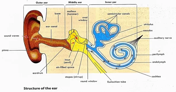
It consists of three main regions: the outer ear, the middle ear and the inner ear.
Outer Ear
The outer ear consists of the visible portion, called the pinna, and the narrow tube-like structure called the ear canal or auditory meatus.
At the end of the canal is the ear drum or tympanum or tympanic membrane. The pinna direct sound through the ear canal to the tympanic membrane.
Middle Ear
The middle ear is air-filled cavity behind the ear drum. It consists of three ear bones called ossicles. These are the malleus (or hammer), incus (or anvil), and stapes (or stirrup). The bones articulate each other and transfer vibrations of the ear drum to the inner ear.
The stapes is connected to the inner ear by the oval window or fenestra ovalis. The middle ear is connected to the pharynx by a narrow tube called the Eustachian tube.
Inner Ear
The inner ear is fluid-filled (perilymph and endolymph). It contains three semi-circular canals which are at right angle to each other.
The canals are borne on the utriculus, which is connected to the cochlea by the sacculus. The ends of semicircular canals are swollen to form an ampulla.
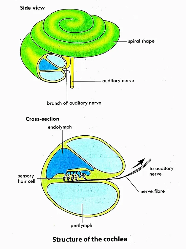
The cochlea, utriculus, sacculus, semi-circular canals contain receptors which are connected and carries impulse to the brain by the auditory nerves.
Mechanism of Hearing
The pinna collects sound waves and pass down the ear canal where they cause the eardrum to vibrate. The vibration of the eardrum sets the ossicles in the middle ear moving against each other. Vibrations of the ossicles amplify the waves which are then passed on to the oval window. The vibration of the oval window causes the fluid in the cochlea to vibrate. The perilymph vibrates and passes onto the endolymph. These waves stimulate the tiny hair cells to produce nerve impulses that travel via the auditory nerve to the brain where they are interpreted as sound.
Mechanism of Balancing
The sensory cells in semicircular canals, utriculus and sacculus are sensitive to the position of the head. The semi-circular canals contain fluid and sensory cells with fine hairs that project into the fluid. When the body loses balance, the fluid in the canals swirl. This stimulates the sensory cells of the ampullae and impulses are sent to the brain (cerebellum) by the auditory nerves. The brain interprets the impulses and sends motor impulses to the appropriate muscles, which reacts to bring the body back to its normal position.
{nextPage}
ENDOCRINE SYSTEM
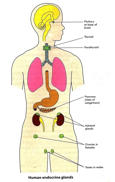
The endocrine system consists of many glands and tissues that secrete hormones into the blood for transport to other tissues and organs. The nervous system and endocrine system both work together to bring about control and coordination in animals.
Types of glands
Endocrine gland: is a ductless gland that secretes hormones directly into the bloodstream.
Exocrine gland: have ducts that carry their secretions to surfaces. Examples include sweat glands, salivary and pancreatic glands, and mammary glands. They are not part of the endocrine system.
Hormones
Hormones are regulatory molecules secreted into the blood by endocrine glands that regulate metabolism of cells, the growth and development of the body parts, emotional behavior and homeostasis.
Characteristics of Hormones
1. Hormones are transported by the blood
2. Hormone produces a specific effect only in its target cells
3. Target cells of a hormone have common specific receptors
4. Hormones are secreted in very small amount
Endocrine Glands
Endocrine glands consist of group of secretary cells surrounded by an extensive network of capillaries which facilitates diffusion of hormone into the blood stream. They are commonly referred to as the ductless glands.
The endocrine glands include;
1. Pituitary glands
2. Thyroid glands
3. Adrenal glands
4. Reproductive glands - the ovaries and the testes
5. The pancreatic islets (islets of Langerhans)
Pituitary Gland
The pituitary gland is attached to the underside of the cerebrum by a stalk. It is often called the “master” endocrine gland because it controls many of the other endocrine glands in the body. The pituitary gland is itself controlled by the hypothalamus.
The pituitary gland consists of the anterior and the posterior pituitary gland
Anterior Pituitary Gland: It secrets six hormones
1. Growth Hormone or Somatotropin (GH): the most abundant hormone synthesized by the anterior pituitary which stimulates the division and growth of body cells. It promotes the synthesis of proteins and other complex biological molecules.
Disorder of growth Hormone
a. Gigantism: occurs as a result of excess concentration of GH during growing years, the individual becomes very tall sometimes nearly 2.5m (8ft) in height.
b. Hypo-pituitary dwarfism: It results when the concentration of growth hormone is low during the growing years and the body growth is limited.
2. Thyroid-stimulating Hormone (TSH): stimulates the thyroid gland to produce thyroid hormone.
3. Adrenocorticotrophic Hormone (ACTH): it controls the secretion of hormones produced by the cortical (outer) portion of the adrenal gland.
4. Prolactin: stimulates milk production by the mammary glands after childbirth.
5. Gonadotrophins: two sex hormones are secreted by the anterior pituitary.
a. Follicle-stimulating hormone (FSH): stimulates production of gametes (ova or spermatozoa).
b. Luteinizing hormone (LH): LH and FSH are involved in secretion of hormones estrogen and progesterone during the menstrual cycle. LH stimulates the interstitial cells of the testes to secrete the hormone testosterone.
Posterior Pituitary Gland
Posterior Pituitary Gland produces two hormones; the antidiuretic hormone and oxytocin.
1. Antidiuretic hormone (ADH): it regulates water reabsorption and blood pressure by increasing the water permeability of the kidney tubules. Disorders of ADH leads diabetes insipidus (excessive water loss in urine), diuresis and dehydration.
2. Oxytocin (OT): Oxytocin is released in large amounts during childbirth. It stimulates and strengthens contraction of the smooth muscles of the uterus during labor.
Thyroid Gland
Thyroid gland is one of the largest endocrine glands in the body. It is situated in the neck in front of the larynx and trachea. Iodine atoms are essential for the formation and functioning of thyroid hormones called thyroxine or T4. It increases the metabolic rate, promote protein synthesis, and enhance neuron function.
Disorders
The disorders may result from hyper-secretion, hypo-secretion and iodine deficiencies
Effects of over secretion (Hyper-secretion)
Effect of under secretion (Hypo-secretion)
Parathyroid Gland: is small glands that are located on the posterior surface of the thyroid gland. It secretes parathyroid hormone (PTH), which regulates blood calcium levels.
Adrenal Glands
Adrenal glands are pair of glands located above the kidneys. Each adrenal gland consists of two distinct portions, an inner adrenal medulla and an outer adrenal cortex.
1. Adrenal medulla: produces the hormone adrenaline (epinephrine) and noradrenaline (norepinephrine). They are released into the blood when stimulated by sympathetic nervous system. They potentiate the fight or flight response by:
b. increasing blood pressure
c. diverting blood to essential organs including the heart, brain and muscles
d. increasing metabolic rate
a. Glucocorticoids: examples cortisol and cortisone. They regulate metabolism and responses to stress. They affect glucose metabolism. The effect of glucocorticoids maintains homeostasis which includes gluconeogenesis and lipolysis.
b. Mineralocorticoids (e.g., Aldosterone): It promotes renal reabsorption of sodium (Na+) and renal excretion of potassium (K+) by the kidney.
c. Sex hormones: Male and female sex hormones similar to those secreted by the ovaries and testes.
Gonads
Gonads are the sex glands; the ovaries and testes. They secrete the sex hormones.
Ovaries: are female gonads, located in the pelvic cavity. They produce two sex hormones.
1. Estrogen: stimulates the development of the female sex organs and secondary sexual characteristics. It also stimulates the thickening of the lining of the uterus for pregnancy.
2. Progesterone: maintains the uterine lining during pregnancy. It prevents ovulation and menstruation. It also stimulates mammary glands for milk production.
Testes: male gonads, produce the hormone testosterone.
Testosterone stimulates the development of the male sex organs, the secondary sexual characteristics and the male sex drive.
Pancreatic Islets
The pancreas contains exocrine and endocrine cells.
1. Endocrine part of the pancreas consists of clusters of small cells called Islets of Langerhans. The beta cells secrete insulin; the alpha cells secrete glucagon.
2. Blood glucose levels are controlled by the opposing actions of insulin and glucagon.
a. Alpha cells secrete glucagon in response to a fall in the blood glucose concentration. The effects of glucagon increase blood glucose levels by stimulating
Disorders (Diabetes mellitus): is caused by the hypo-secretion of insulin. It is characterized by excessively high levels of glucose in the blood.
Differences between hormonal coordination and nervous coordination in humans
|
Hormonal |
Nervous |
|
Transmission is chemical |
Transmission is electrical and
chemical |
|
Slow
transmission |
Rapid transmission |
|
Response is wide
spread |
Response localized |
|
Target
organs receive response |
Effector organs receive
response |
|
Long lasting response |
Short-lived response |
|
Pathway
not specific through blood |
Pathway specific through
nerve fibers |
Get Your Free PDF!
Download our comprehensive guide to
THE NERVOUS SYSTEM AND ENDOCRINE SYSTEM IN HUMANto boost your knowledge.
Click the button above to get instant access to the PDF.
Related Post on Biology Topics
Click Here for WAEC/ SSCE/ WASSCE/ NOVDEC Past Questions and Answers on Coordination and Control



%20%20brain%20with%20labels.webp)



.webp)

%20and%20its%20correction.webp)
%20and%20its%20correction.webp)
.webp)
