THE SKELETAL SYSTEM AND MOVEMENT IN MAMMALS
Objectives
This blog post provides readers with the following objectives. The reader will be able to:
MAMMALIAN SKELETON
Skeleton is the hard parts of an animal that forms the framework for the body. Mammalian skeleton is composed of three types of organs; bones, cartilages and ligaments, tightly joined together.
Types of skeletons
1. Hydrostatic Skeleton: is found in soft body animals e.g. hydras, flatworms, annelids.
2. Exoskeletons: are hard structures found outside the body of an animal. They enclose and protect soft tissues and organs of the body. E.g. beetle, cockroach, snails, crab etc.
3. Endoskeletons: are rigid, jointed and lie within the body of an animal. They are composed of living tissues (bone and cartilage). E.g. fish, toad, lizard, birds, mammals.
Functions of the Mammalian skeleton
1. Red blood cells are manufactures in the bone marrow.
2. Forms the rigid support of the body
3. Provides point for muscles attachment
4. Provides series of levers for movement
5. Gives shapes to the body
6. Protects the delicate internal organs
7. It stores minerals such as calcium and phosphorus
General Plan of Mammalian Skeleton
The bones of the skeleton consist of two groups:
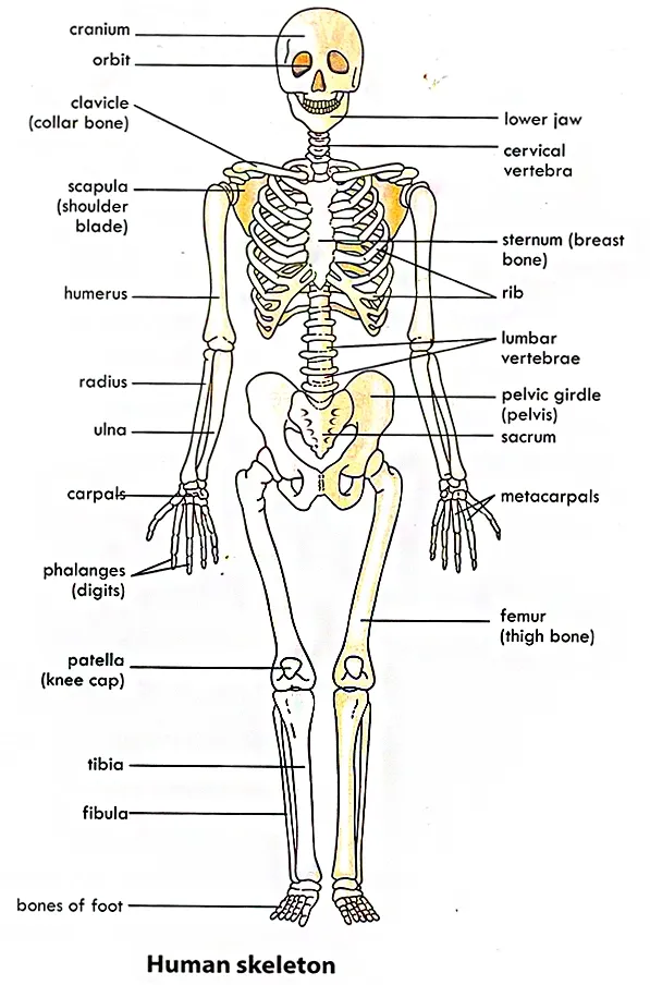
1. Axial Skeleton made up of the skull, vertebral column, ribs, and sternum.
2. Appendicular Skeleton made up of forelimbs and hind limbs
Skull
The skull protects the brain and sense organs. Cranial bones are flat bones that surround and protect the brain.
Vertebral Column
The vertebral column (back bone) consists of a series of irregular
bones called vertebrae, separated from each other by fibro-cartilaginous called
intervertebral discs.
The vertebrae are held together by ligaments which prevent their dislocation. Between
the vertebrae are opening called intervertebral foramina that allow
passage of spinal nerves.
There are five different types of vertebrae in human. They are named according to location:
General Structure of Vertebrae
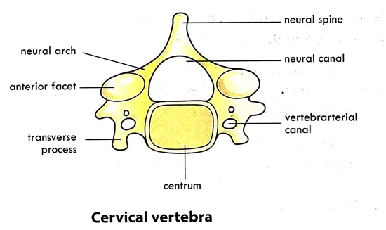
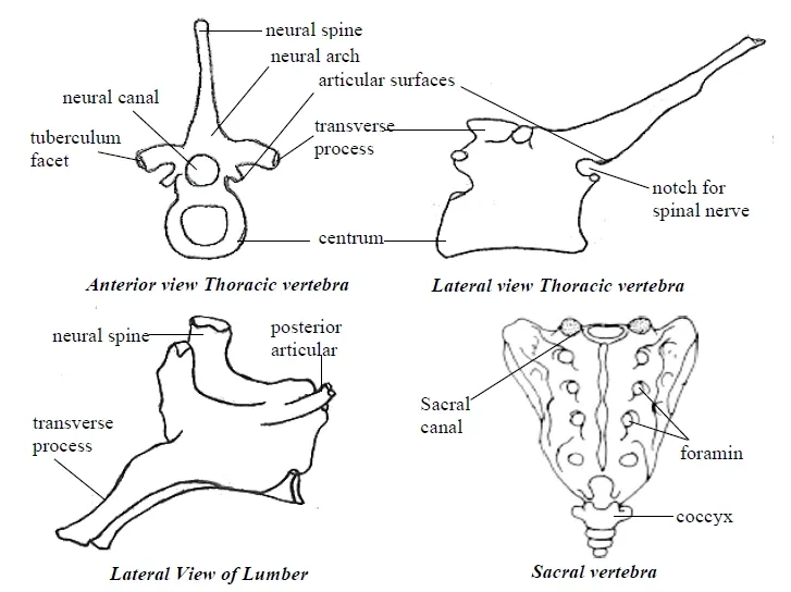
1. Centrum: it forms the body of the vertebra. The centrum fits into the intervertebral dics on both sides. The centrum carries the neural arch and neural spine.
2. Neural arch: is attached to the posterior surface of the body. It forms an arch over the centrum and surrounds the neural canal. The neural arch extends dorsally upwards to form neural spine. The neural arch gives off transverse processes; the anterior process called prezygapophyses and those at the posterior part are called post-zygapophyses.
3. Neural canal: is a central canal which serves as passageway for the spinal cord.
Cervical Vertebrae
The cervical vertebrae are the smallest of the bones. There are seven cervical vertebrae in the neck region. They have two vertebral canals through which blood vessels of the neck pass. The first and the second cervical vertebrae are peculiar in shape.
Characteristics of cervical vertebral
1. centrum is small and oblong in shape
2. neural canal is large
3. presence of a vertebraterial canal or foramen in each transverse process
4. reduced or short neural canal
Atlas
The atlas is the first cervical vertebra. It is almost ring like. It provides up and down or nodding movement to the skull.
Characteristics
1. absence of centrum
2. large neural canal
3. short rounded neural spine
4. flat and broad transverse process
5. presence an anterior and posterior arch.
6. it has articular facets which articulate with the oval occipital condyles of the skull to form atlanto-occipital joint
Axis
The second cervical vertebra is termed as axis. Its centrum bears an odontoid process, which allows side to side or turning movement of the skull.
Characteristics
The atlas and axis are specialized to support the head and allow it to nod “Yes” and shake “No”
Function of cervical vertebra
1. form the framework of the neck and support the head region.
2. provides surfaces for the attachment of the neck muscles
3. vertebrate rial canal provides pathway for blood vessels and nerves in the neck region
Thoracic Vertebrae
Thoracic vertebrae are larger than cervical vertebrae. The twelve vertebrae in the thoracic region constitute the thoracic vertebrae. Each thoracic articulate with a pair of ribs using its capitular and tubercular facets.
Characteristics
1. have heart-shaped centrum with articular facets for attachment of the ribs.
2. neural arch is relatively small.
3. neural spine is long and is directed downwards
4. thick and strong transverse processes articular facets which support the ribs
Lumbar Vertebrae
Lumbar Vertebrae are located in the abdomen. They are the largest vertebrae of the vertebral column. Lumbar vertebrae have projections for attachment of powerful back muscles.
Characteristics
The sacral vertebrae
It consists of five sacral vertebrae, which are fused into a single bone called sacrum. The wedge-shaped sacrum provides a strong foundation for the pelvic girdle.
Characteristics
Coccygeal or Caudal Vertebrae
These are 4 in number and occur in the vestigial tail. They are very small and fused to form a curved, triangular bone called the coccyx or tail bone. The first vertebra of the fused coccyx attached to the sacrum by ligaments. The coccyx flexes anteriorly, acting as a shock absorber, when a person sit. It also supports the tail.
Characteristics
1. Absence of neural canal neural arch, neural spine
2. Lack transverse process
3. Thick centrum
Caudal vertebrae progressively decrease in size from the base of the sacrum to the tip of the tail.
Functions of Vertebral Column
1. The vertebrae enclose and protect the spinal cord
2. Support the skull and allow for its movement
3. Articulate with the rib cage
Rib Cage or Thoracic Cage
1. It protects vital organs (heart, lungs and blood vessels)
2. It supports the pectoral girdle
3. It involves in respiration (breathing)
4. Bone marrow in certain rib bones produce blood cells
{nextPage}
Appendicular Skeleton
The appendicular skeleton is divided into
1. pectoral girdle and the pelvic girdle
2. upper (Fore) limbs and lower (Hind) limbs
Pectoral (Shoulder) Girdle
Functions
1. clavicle holds the scapula in position
2. supports the arm
3. scapula provides surfaces attachment of shoulder muscles
4. the acromion process articulate with the clavicle to allow sliding movement
5. glenoid cavity of scapula articulates with head of humerus to allow movement in any plane
Pelvic (Hip) Girdle
Pelvic girdle or hip girdle is formed by a pair of hip bones, each called innominate bone. Each large, irregularly shaped hip bones consist of three separate bones; the ilium, ischium and pubis. In adults these bones are firmly fused and their boundaries are indistinguishable. At the point of fusion of the ilium, ischium and pubis is a deep hollow called acetabulum on the lateral surface of the pelvic. The acetabulum receives the head of the femur at this hip joint. The pubis and ischium enclose a large hole called obturator foramnen. The deep basin-like structure formed by the hip bones together with the sacrum and coccyx is called the bony pelvis.
Functions
3. ilium has large surface area for attachment of the hip muscles
4. Acetabulum articulates with head of femur to form the hip joint
Upper Limb (Fore-Limb)
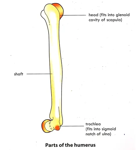
Humerus (upper arm): is the bone of the arm. It articulates with the scapula at the shoulder joint and with the radius and ulna (forearm bones) at the elbow. It consists of three sections:
1. Proximal part consisting of a rounded head, a narrow neck, and two short processes called tuberosities or t Cylindrical body or shaft
2. Distal part consisting of two condyles, processes (trochlea & capitulum), and fossae
Functions of Upper limbs
1. The smooth rounded head fits into the glenoid cavity of the scapula.
2. The tuberosity is a site for muscles attachment.
3. The trochlea articulates with the sigmoid notch of the ulna.
4. The capitulum articulates with the radius.
5. The supratrochlear foramen provides a passage for blood vessels and nerves
{nextPage}
Bones of the wrist and hand
Lower Limb (Hind Limb)
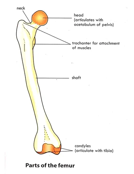
Femur: is the longest, heaviest and the strongest bone in the body. The
proximal rounded head of the femur articulates with the acetabulum of the pelvic
girdle to form hip joint. The constricted region supporting the head is called
the neck.
The body
(shaft) has several features for muscle attachment. The distal end of
the femur is expanded for articulation with the tibia.
Tibia and Fibula: these are the bones of the leg. The tibia articulates proximally with the femur at the knee and distally with the talus of the ankle. It also articulates both proximally and distally with the fibula.
{nextPage}
Joint and Muscles
Joints or Articulations are sites where two or more bones meet. Joints give skeleton mobility and also hold bones together
Classification of Joints
Joints are classified based on the material binding the bones together.
1. Fibrous (or Fixed) Joints: are immovable. The articulating bones are joined by fibrous tissues. No joint cavity is present. E.g. Sutures of the skull.
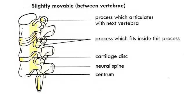
2. Cartilaginous (or Slightly Movable) Joints: are slightly movable. The articulating bones are united by cartilage. They lack a joint cavity. E.g. intervertebral joints.
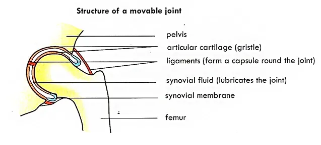
3. Synovial (or Movable) Joints: are freely movable. The articulating bones are separated by a fluid-containing-joint cavity. This arrangement, permits freely movement synovial joints. E.g. pivot joint, hinge joint.
Types of Synovial Joints
1. Plane (or Gliding) joints: allow only short slipping and gliding movements e.g. the intercarpal and intertarsal joints.
2. Hinge joints: the movement is along a single plane and is similar to that of a mechanical hinge. E.g. knee joint and elbow joint.
3. Pivot joints: allow rotation of the bone around its own axis or over the other. E.g. is the joint between the atlas and the axis.
4. Ball and socket joints: allow a wide range of movement. E.g. the joints at the shoulder and hip
General Structure of Synovial Joints
Muscle tissues are made up of elongated, elastic cells called muscle fibers. The elongated and elastic feature helps muscle to contract and relax. Contraction and relaxation of muscles help in movement. Muscles usually work in pairs. When one contracts the other relaxes and vice versa. Pairs of muscles that work like this are called antagonistic muscles. The biceps and triceps muscles of the arm are an example of an antagonistic pair.
{nextPage}
Types of Muscle
The 3 types of muscle tissue are skeletal, cardiac and smooth.
Skeletal, Voluntary or Striated Muscle
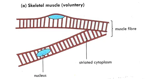
It is attached to and covers the bony skeleton. Skeletal muscle
cells or fibers are long, threadlike, with dark cross-markings or stripes called
Striations. Each fiber contains many nuclei located just beneath
its cell membrane (sarcolemma). Skeletal muscle is called
voluntary muscle because it is the only type subject to conscious control.
The cytoplasm (sarcoplasm) of the fibers contains:
1. Bundles of myofibrils which consist of fine filaments (or sarcomeres) of contractile proteins including actin and myosin.
2. Many mitochondria that generate energy (ATP) from glucose by cellular respiration.
3. Glycogen, a store carbohydrate which is broken down into glucose when required.
4. Myoglobin, a unique oxygen-binding protein that stores oxygen within muscle cells.
For more on muscle fiber types, refer to this article
For more details, visit:
Cardiac Muscle Tissue: Structure and Function
Cardiac muscle is only found in the wall of the heart. It is striated, but it is not voluntary. The muscle fibers are branched and interconnected to form complex networks. The fibers are composed of cells which are joined end to end. Each cell has a single nucleus. Each cell is attached another cell at a specialized intercellular junction called an intercalated disk. A wave of contraction spreads from cell to cell across the intercalated discs.
Function
Contraction and Coordination: A wave of contraction spreads from cell to cell across the intercalated discs. This coordinated contraction ensures that the heart pumps blood efficiently throughout the body.
For more detailed information on cardiac muscle tissue, visit these resources:
- NCBI: Cardiac Muscle Tissue Overview
- Khan Academy: Cardiac Muscle Structure
- Mayo Clinic: Cardiac Muscle Function
Smooth Muscle Tissue
Smooth muscle is found in the walls of the hollow internal organs
such as the stomach, urinary bladder, blood vessels and the respiratory
passages. It is called smooth because its cells lack striations. It is described mostly as visceral,
nonstriated and involuntary. Contractions of smooth
muscle fibers are slow and sustained. The cells are spindle-shaped
with only one central nucleus.
The cells are shorter than those of the skeletal muscle.
Function 0f smooth muscle
1. Movement and Control of Substances
2. Regulation of Blood Flow and Pressure
3. Control of Airflow in Respiratory Tract
For more detailed information on smooth muscle tissue, visit these resources:
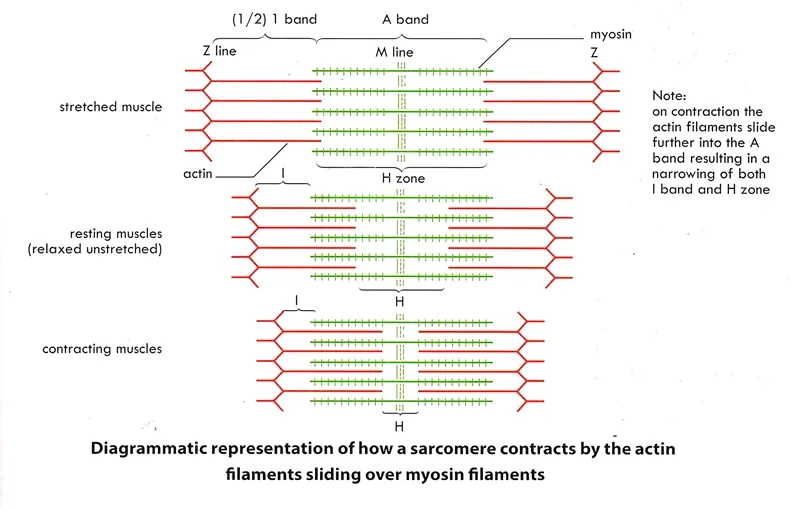
The Sliding-Filament Model of Muscle Contraction
The sliding filament theory of muscle contraction is the binding of
myosin to actin, forming cross-bridges that generate filament
movement. Muscles contract when sarcomeres shorten. The thin and thick
filaments that compose sarcomeres, slide past one another, causing the
sarcomere to shorten while the filaments remain the same length.
Movement of the Fore Limb
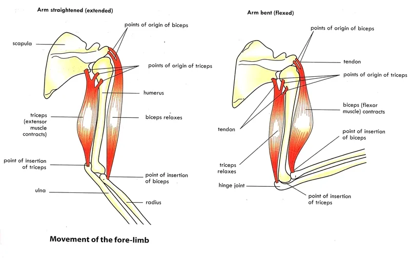
A single muscle is fat in the middle and tapers towards the ends (i.e., the origin and the insertion). The middle part, which gets fatter when the muscle contracts, is called the belly of the muscle.
Skeletal muscles usually work in pairs. When
one contracts the other relaxes and vice versa. Pairs of muscles that work like
this are called antagonistic muscles. Example of antagonistic muscle
is the biceps and triceps in the upper forearm.
Together they bend and straightened the forearm.
The origin of the biceps is attached to the scapula (the bone that moves the least when muscle contract) while the insertion is attached to the radius (movable part). When the biceps contracts (and the triceps relaxes) the lower forearm is raised and the angle of the joint is reduced. This kind of movement is called flexion. When the triceps is contracted (and the biceps relaxes), the angle of the elbow increases (i.e., straightening the lower arm). The term for this movement is extension.
References
1. Skeletal System Overview
- NIH Osteoporosis and Related Bone Diseases National Resource Center offers comprehensive information on bone biology, development, and disorders.
- Khan Academy offers free educational materials on the skeletal system, including videos and articles on bone anatomy and physiology.
- National Institute of Arthritis and Musculoskeletal and Skin Diseases (NIAMS) provides comprehensive information on bone health and osteoporosis, which is crucial for understanding the skeletal system.
- Encyclopædia Britannica offers detailed articles on the skeletal system in humans and other animals, including the structure and function of bones.
Get Your Free PDF!
Download our comprehensive guide on
THE SKELETAL SYSTEM AND MOVEMENT IN MAMMALS.
Download PDFClick the button above to get instant access to the PDF.
Related Post on Biology Topics
Click Here for WAEC Past Questions and Answers on Movement in Mammals










%20types%20of%20joint%20in%20skeleton.webp)
.webp)
%20types%20of%20joint.webp)
.webp)

.webp)
