Understanding Nucleic Acids and Protein Synthesis: A Comprehensive Guide
Objectives
This blog post provides readers with the following objectives. The reader will be able to:
NUCLEIC ACIDS
Nucleic acids are large biological molecules, essential for all known forms of life. It forms the genetic material of living organisms. Nucleic acids are made from monomers known as nucleotides. There are two types of nucleic acids: deoxyribonucleic acid (DNA) and ribonucleic acid (RNA).
DNA
DNA is present in the nucleus of all cells and it contains genetic information that allows living things to function, grow and reproduce. It carries genetic information from one generation to another. The units of inheritance called genes are actually sections of the DNA molecule.
Structure of DNA
DNA is a large molecule made up of a long chain of sub-units or monomers called nucleotides. Each nucleotide is made up of;
1. pentose sugar called deoxyribose
The bases: nitrogen base is grouped into two;
1. Purines: made up of Adenine and Guanine.
2. Pyrimidine: made up of Cytosine and Thymine.
The deoxyribose, the phosphate and one of the bases combine to form a nucleotide. The combination of a base and a sugar without phosphate group is called nucleoside.

DNA Packaging
DNA in eukaryotic cells is organized into structures called chromosomes. To fit within the nucleus, DNA is wrapped around histone proteins, forming nucleosomes. This higher-order packaging is essential for regulating gene expression and ensuring efficient DNA replication.
For more on DNA packaging, visit Chromosome Organization and Packaging.
The Importance of DNA Structure
Understanding the structure of DNA has profound implications for various fields:
- Genetics: The double helix model elucidates how genetic information is stored, replicated, and passed on to offspring.
- Medical Research: Insights into DNA structure have led to advancements in genetic disorders, gene therapy, and personalized medicine.
- Forensic Science: DNA profiling is a powerful tool in criminal investigations and paternity testing.
Explore how DNA Research Transforms Medicine and its applications in real-world scenarios.
DNA Replication
DNA replication occurs before cell division.
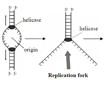
Replication bubble forms at a point in the DNA called the origin of replication. The enzyme helicase unwinds the DNA molecule by breaking hydrogen bonds between the bases. The top half of the replication bubble looks like an upside down "Y". This area is called a replication fork. The separated strands serve as templates for the synthesis of new DNA.
DNA polymerase synthesizes new DNA strand complimentary to the separated strands. The direction of synthesis is 5' to 3'. One side of the strand is synthesized continuously whiles the other strand is synthesized in fragments. The strand that is synthesized continuously is called the leading strand. The strand that is synthesized in fragments is called the lagging strand. The fragments are called Okazaki fragments.
Ligase catalyzes the formation of covalent bonds between the Okazaki fragments.
RIBONUCLEIC ACID (RNA)
Ribonucleic acid (RNA) is made up of a long strand of nucleic acids similar, but not identical to DNA. It occurs in both the nucleus and cytoplasm. It consists of small sub-units called nucleotides which are composed of:
1. Ribose pentose sugars (C5H10O5)
2. Phosphates
3. Nitrogenous base consisting of two purines (Adenine and Guanine) and two pyramidine (Cytosine and Uracil). Thymine is replaced by Uracil
The base pairing rule Adenine (A) pairs with Uracil (U) and Guanine (G) pairs with Cytosine (C). RNA is single-stranded molecule and has a much shorter chain of nucleotides.
Types of RNA
There are three types; transfer RNA (tRNA), messenger
RNA (mRNA), and ribosomal RNA (rRNA).
Messenger RNA (mRNA)
mRNA constitutes 3 to 5% of the total RNA. Its single stranded molecule formed on a single strand of DNA in process called transcription.
mRNA carries information from DNA to the ribosome; the sites of protein synthesis in the cell. It serves as the template for the synthesis of a protein.
Its size depends on the size of the protein for which it codes. It’s made up of several thousands of nucleotides.
The base sequence of mRNA is in triplets each of which is called a codon.
Transfer RNA (tRNA)
tRNA constitutes 15% of the total RNA. It is a small molecule, containing 73-93 nucleotides.
It carries amino acids to the site of protein synthesis, (the ribosome). Many of the bases in the chain pair with each other forming sections of double helix.
The unpaired regions form 3 prominent loops.
Each tRNA has a three-base sequence at one loop called the anticodon which binds to a complementary triplet codon on the mRNA. Each kind of tRNA carries (at its 3′ end) specific amino acids.
The 3′ end of every tRNA molecule ends with nucleotides containing CCA bases.
Ribosomal RNA
rRNA constitutes about the 80% of the whole RNA present in eukaryotic cell. rRNA combines with proteins to forms the ribosome which is the site of protein synthesis. rRNA is the catalyst for formation of the peptide bond.
The ribosome structure is composed of 2 subunits. A small and a large subunit each of which consists of rRNA and a small quantity of proteins.
Functions of RNA
- Protein Synthesis: RNA is directly involved in translating the genetic code from DNA into proteins, which perform essential functions in the body.
- Gene Regulation: Certain RNA molecules can regulate which genes are turned on or off in a cell.
- Catalysis: Some RNA molecules, known as ribozymes, have catalytic activity and can speed up biochemical reactions.
- Genetic Information: In some viruses, RNA carries genetic information instead of DNA.
Importance in Research and Medicine
RNA research has led to significant advancements in medicine, including the development of RNA-based vaccines and therapies for various diseases. The study of RNA interference (RNAi) has opened new pathways for gene silencing, providing potential treatments for genetic disorders.
Difference between DNA and RNA
|
DNA |
RNA |
|
Found in the nucleus |
Found in the nucleus and cytoplasm |
|
Pentose sugar is deoxyribose(C5H10O4) |
Pentose sugar is ribose (C5H10O5) |
|
Double stranded |
Single stranded |
|
Can replicate |
Cannot replicate |
|
Bases are adenine, guanine, cytosine and thymine |
Bases are adenine, guanine, cytosine and uracil |
|
Has very high molecular weight and contains long chain nucleotides |
Has relatively small molecular weight and consist of few
nucleotides |
|
Base pairing rule; adenine pairs with thymine and cytosine pairs
with guanine |
Base pairing rule; adenine pairs with uracil and cytosine pairs
with guanine |
|
It controls heredity and protein synthesis |
It is involved in protein synthesis |
For more detailed information on RNA, visit National Human Genome Research Institute's RNA overview
PROTEIN SYNTHESIS
Protein synthesis occurs on the ribosome in the cytoplasm. Protein synthesis involves amino acids and RNA, catalyzed by enzymes. It requires two steps: transcription and translation.
Transcription
Transcription is the process where the information in a DNA strand is transferred to an RNA molecule to form messenger RNA (mRNA). The RNA is called messenger RNA because it carries the 'message' or genetic information from the DNA to the ribosomes, where the information is used to make proteins. Transcription occurs in the nucleus.
Steps Involved in Transcription
1. DNA is transcribed by an enzyme called RNA polymerase.
2. RNA polymerase binds to DNA and unwinds the double helix.
3. One strand of DNA serves as the template for RNA synthesis. Thus,
the coded information of the DNA is copied across or transcribed into an mRNA.
4. The strand that serves as the template is called the antisense
strand. The strand that is not transcribed is called the sense
strand.
5. The mRNA molecule leaves the nucleus and enters the cytoplasm.
Translation
Translation is the process where ribosomes synthesize proteins using the information on mRNA produced during transcription. The mRNA attaches itself to a ribosome in the cytoplasm. The base sequences of nucleotides on the mRNA strand are in triplet (called codon), which are instructions for the synthesis of protein. Each code or codon is specific for one amino acid.
Steps in Translation
1. Ribosome and first tRNA comes together at AUG start codon on mRNA.
2. In eukaryotes, first or initiator tRNA carries methionine (met). The
initiator tRNA having met at its free end, pairs with the start codon on mRNA.
3. A second tRNA molecule transports the next amino acid to the
ribosome. The second tRNA with its amino acid binds to the codon next to the
start codon.
4. A peptide bond is formed between the two adjacent amino acids.
5. The bond between the first tRNA and its amino acid (met) breaks.
tRNA is released from the ribosome.
6. rRNA catalyzes the peptide bond formation and breaks the bond
between amino acid and tRNA.
7. The ribosome moves along the mRNA to another codon for another tRNA
molecule.
8. Third tRNA transports other amino acids base on it anticodon. A
second peptide bond is formed between the second and third amino acids.
9. The bond between the second tRNA and its amino acid breaks and it
detaches itself from the ribosome.
10. The ribosome moves along the mRNA strand, the cycle repeats itself and
the synthesis continues until it reaches the stop codon or non-sense codon (UAA, UAG
or UGA).
11. There are no tRNA molecules with anticodons for STOP codons.
12. A release factor binds to a stop codon causing the polypeptide chain
to be released and causing the ribosome and mRNA to dissociate.
Importance of Protein Synthesis
Protein synthesis is essential for:
- Cell Growth and Repair: Producing new proteins for cell structure and function.
- Enzyme Production: Synthesizing enzymes that catalyze biochemical reactions.
- Regulation: Producing hormones and other regulatory proteins.
For a deeper dive into protein synthesis, check out Nature's guide to protein synthesis.
Genetic Code
Genetic code is the set of rules by which information encoded within
genetic material (DNA or mRNA sequences) is translated into proteins by living
cells. The genetic code by which DNA stores the genetic information consists of
"codons" of three nucleotides. The functional segments of DNA which
code for the transfer of genetic information are called genes.
With four possible bases, the three nucleotides (codon) can give 43 = 64 different genetic codes. 61 codons code for amino acids. Three codons UAA, UAG and UGA are describe as stop codons. They do not code for any amino acids.
The genetic codes for each
amino acid
|
RNA triplet |
Amino Acid |
Abbreviation |
RNA triplet |
Amino Acid |
Abbreviation |
|
AAA AAG |
Lysine |
Lys |
GAC GAU |
aspartic acid |
Asp |
|
AAC AAU |
Asparagine |
Asn |
GCA GCC |
Alanine |
Ala |
|
ACA ACC |
Threonine |
Thr |
GGA GGC |
Glycine |
Gly |
|
AGC AGU |
Serine |
Ser |
GUA GUC |
Valine |
Val |
|
AUA AUG |
Methionine |
Met |
UUA UUG |
Leucine |
Leu |
|
AUC AUU |
Isoleucine |
Ile |
UAC UAU |
Tyrosine |
Tyr |
|
CAA CAG |
Glutamine |
Gln |
UCA UCC |
Serine |
Ser |
|
CAC CAU |
Histidine |
His |
UGC UGU |
Cysteine |
Cys |
|
CCA AGG |
Proline |
Pro |
UGG |
Tryptophan |
Try |
|
CGA CGC |
Arginine |
Arg |
UUC UUU |
phenylalanine |
Phe |
|
UAA UAG |
(stop) |
|
|
|


.webp)


